
A Review of the Dinoflagellates and Their Evolution from Fossils to Modern

1
British Geological Survey, Keyworth, Nottingham NG12 5GG, UK
2
Geological Survey of Canada, P.O. Box 1006, Dartmouth, NS B2Y 4A2, Canada
3
C.N.R.S. (Centre National de la Recherche Scientifique), Laboratoire Arago, Sorbonne University, 66650 Banyuls-sur-Mer, France
4
Marine Biological Association of the UK, Plymouth PL1 2PB, UK
*
Author to whom correspondence should be addressed.
J. Mar. Sci. Eng. 2023, 11(1), 1; https://doi.org/10.3390/jmse11010001
Received: 28 October 2022
/
Revised: 7 December 2022
/
Accepted: 8 December 2022
/
Published: 20 December 2022
(This article belongs to the Special Issue Marine Phytoplankton and Their Evolution)
Abstract
:Molecular clock and biogeochemical evidence indicate that the dinoflagellate lineage diverged at around 650 Ma. Unequivocal dinoflagellate cysts/zygotes appeared during the Triassic. These biotas were badly affected by the end-Triassic extinction and recovery from this was relatively slow. During the early Middle Jurassic, the family Gonyaulacaceae underwent an explosive diversification event and taxonomic richness steadily increased throughout the rest of the Jurassic. The entire Cretaceous also recorded increases in diversity. This trend reversed during the Oligocene, probably caused by global cooling. Marine cyst-forming peridiniaceans declined substantially through the Oligocene and Neogene, but protoperidiniaceans continued to diversify. Modern taxa, as evidenced by the molecular tree, comprise three major clades: the first two are composed largely of parasitic forms, marine alveolates of unknown identity and the Syndiniales; free-living dinoflagellates form the third clade, which diverges rapidly and bears short branch lengths with no real support for branching order. This suggests that morphological divergence preceded molecular divergence because, as the fossil record indicates, major groups appeared at different ages. Unique features of the dinoflagellates helped the group take on a predominant role in the marine phytoplankton. Living in marine or fresh water, dinoflagellates have demonstrated innovative capacities that have enabled them to live among the phytoplankton or benthos as autotrophic, heterotrophic, mixotrophic free-living organisms or symbiotic and/or as parasitic forms.
1. Introduction
Dinoflagellates are an important member of the microplankton, especially because they have both pigmented and non-pigmented species, which means that they have various ecological roles, for example as autotrophs and heterotrophs. Thus, the group plays a prominent role in the ecology of aquatic environments. In the current classification, 550 genera of dinoflagellates are recognised, containing approximately 6000 described species. Living species number ca. 3794 species (www.alagebase.org, accessed on 6 December 2022). Taylor [1] noted the immense diversity of the dinoflagellates. Traditionally, dinoflagellates have been divided into armoured and naked forms. The armoured dinoflagellates possess distinctive plates, composed of cellulose-like material, inside flattened vesicles underneath the plasma membrane. Such vesicles are shared with related groups, such as the ciliates and are called alveolae generally and amphiesmal vesicles among dinoflagellates. Following the phylogeny of Bachvaroff et al. [2], for example, dinoflagellates belong to alveolates and can be autotrophic (about half of the species are photosynthetic), heterotrophic, parasitic and/or mixotrophic, or symbiotic, plasmodial, or be organised as “pseudo-multinucleated” protists [3].
Dinoflagellates all live in aquatic environments, be it salt, brackish or fresh water. The composition and original organization of their nucleus (dinokaryon), with condensed chromosomes and chromatin, as well as a distinctive mitosis, are additional distinct features. Their mitotic apparatus (except in the Syndiniales and Oxyrrhis sp.; see below) is different from that in all other eukaryotes and has also been considered as an evolutionary marker (Figure 1) [4].
The authorities of all fossil-based dinoflagellate genera, species and cysts mentioned in this contribution are listed in Appendix A. Authorities for living species can be found in Algaebase. We follow the classification in Algaebase throughout.
2. Feature of the Living Cells and Phylogeny of Living Dinoflagellates
2.1. Nuclear Characters
Within the dinokaryon, chromosomes are maintained in a quasi-permanent condensed state in most dinoflagellate species. Chromosome separation is accomplished by an extranuclear microtubular mitotic spindle located in cytoplasmic channels that pass through pores in the nuclear membrane during mitosis [5,6]. The extranuclear spindle manipulates the chromosomes to achieve cell division.
Edouard Chatton was the first researcher to take a detailed interest not only in the description of the external morphology of many dinoflagellates, but also in their life cycle, cytology, and dino- and syndinian mitosis [7,8,9]. For example, Prorocentrum micans has a nucleus filled with hundreds large chromosomes (10 µm long by 1 µm wide). The nucleus is surrounded by large plastids. Each chromosome is superhelically twisted by nucleofilaments [10], which are devoid of histones and nucleosomes but are rich in a particular base, hydroxymethyluracyl [11].
Dinoflagellates can reproduce either by vegetative binary reproduction or by sexual reproduction (see Table 1, Figure 2), the latter often occurs when physico-chemical conditions are unfavourable, such as at the end of a bloom. In armoured dinoflagellates, cells pair up and a tube usually develops to connect the mating pairs through which genetic material is exchanged [12,13]. Shortly after nuclear fusion, their chromatin is animated by a movement (chromatic cyclosis) for several minutes while male and female genetic materials mingle and the chromosomes lose their regular helical condensed structure. This cyclosis, discovered by Pouchet [14] in two species of Ceratium, was clarified by Biecheler in six species of Peridinium [15]. The planozygotes (swimming zygotes) containing diploid DNA undergo meiosis, result in the production of haploid vegetative cells [12]. Subsequent observations have shown that nuclear cyclosis occurs directly before the first meiotic division and that the organization of chromosome nucleofilaments architecture was modified [13]: after a high decondensation and during the rotation of the chromatin, chromosomes are very stretched, showing a visible axis, multiple loops and figures of crossing over. During the first meiotic division, relaxed chromosomes begin to divide.
2.2. Phylogenetic Analysis
A RAxML bootstrap (BT) 18S rRNA tree is presented in Figure 3. The tree is rooted by three metazoan outgroups and several apicomplexans. Clades are collapsed to show major divergences in the dinoflagellates but the tree in the supplemental data shows each clade fully open so that all species can be seen. Each collapsed clade is colour-coded as to whether its members are entirely naked (green) or armoured (blue) or pink if it is mixed with both naked and armoured members. What is immediately noticeable is that there is no absolute division between naked and armoured dinoflagellates, although naked dinoflagellates are in a basal position. There are three major clades. The first two are primarily composed of syndianialean species and correspond to two of the five dinoflagellate classes as recognised in Algaebase. These divergences are followed by sequential divergences of Heterodinium and Katodinium glaucum. The final third large clade contains the majority of the dinoflagellates. Each clade is discussed in detail with innovative features that distinguish that clade. Each of the smaller clades within this third large clade are supported by bootstrap (BT) values greater than 85% but the branching order is not well supported except where noted in the text because of the rapid diversification of the group.
2.2.1. Clade 1
The first divergence (Figure 3 and Figure 4) contains two clades: a naked one with Amphidinium spp. and a naked one that contains uncultured alveolates from the so-called group 1 alveolates and two parasitic dinoflagellate genera (Euduboscquella and Ichthyodinium), which fall into the Syndiniales group 1 classification [16], Kim et al., 2008. These two genera parasitise tintinnids and fish eggs, respectively. Two sequences of Karlodium veneficum are included in this clade and are likely derived from parasites within the host cell because all other sequences of members of the Kareniaceae are in a different monophyletic clade. A GenBank Blast of the two sequences labelled as Karlodium veneficum shows that they are clearly similar to other marine alveolates in this region of the tree and thus the name in GenBank is taxonomically incorrect. The genus Euduboscquella includes intracellular parasitic dinoflagellates that divide only after exiting the host cell. They show characteristic features during the trophic growth stage [16] Kim et al., 2008. The division products from a single infection produce either motile dinospores or nonmotile spores. These dinoflagellates form the class Ellobiophyceae.
The basal position of Amphidinium and its reoccurrence in a basal position in other parts of the tree would suggest that the ancestral dinoflagellate may have had apical flagella. It would seem logical that, as the girdle region developed and the two flagella became more specialised, flagellar insertion would have begun in a more apical position and (in an evolutionary sense) moved toward the centre of the cell, rather than the reverse. If an initial apical insertion is correct, then the small episome seen in Amphidinium and related species is not a derived state but an ancestral one.
2.2.2. Clade 2
The second divergence (clade 2) contains both naked and armoured dinoflagellates (Figure 5). The Syndiniales Group 2 parasites and Hematodinium represent naked dinoflagellates. Hematodinium is in the sister clade to group 2 Syndiniales, and this genus parasitises decapod crustaceans. So this non-planktonic host may be significant in the relationships between dinoflagellates that parasitise other plankton versus those that parasitise benthic hosts. In comparison, the syndinialean genus, Amoebophrya, found in syndinean clade II, produces a single type of spore and undergoes nuclear division inside the host cell [16]. There is some evidence that Syndiniales Group 1 species can infect more than one host and more than one Group 1 spp. may infect the same host species, whereas studies with Group 2 species, specially Amoebophrya, have suggested host parasite specificity [16]. The vast diversity of environmental clone library sequences suggests that both groups of Syndiniales contain a diverse array of potential parasite species. These dinoflagellates form the class Syndiniophyceae.
The armoured clade is basal to the naked clade and is represented by the peridinialean genus, Pentapharsodinium, which is a polar to equatorial euryhaline genus. The basal position of the thecate forms in this clade suggests that these parasites had a free-living thecate ancestor.
2.2.3. Innovative Features of the First Two Clades
These early divergent groups, which form two of the five classes in the dinoflagellates (see www.Algaebase.org, accessed on 5 December,2022) may have taken advantage of their mixotrophic life style, by being heterotrophic during their parasitic stages in their hosts and being autotrophic, using photosynthesis, during the free-living stages as reproductive dinospores. Chatton [8] noted that such forms are present in all sorts of ecological niches: intracytoplasmic and intranuclear parasites of protozoans; symbionts of protozoans and metazoans; intestinal, coelomic, or haemolymphatic parasites of metazoans; and external parasites of metazoans (Figure 6). The rapid reproduction of the parasitic stages means that these dinoflagellates can very quickly increase their cell numbers to dominate or control a bloom situation of the planktonic host cells, or decimate benthic host populations.
Chatton considered syndinian mitosis a unique type of mitosis within the dinoflagellates and described it in detail [7,8] from several species parasitizing various copepod crustaceans. Subsequently, TEM observations demonstrated the distinctive nature of this mitotic system: the 5 compacted chromosomes are attached to the extranuclear microtubular mitotic spindle through the nuclear membrane by means of kinetochores via a large cytoplasmic channel. They are connected to the centrosome region containing two centrioles (Figure 6a,b and Figure 7) [17,18]. Of particular interest is the appearance of histone-like basic nuclear proteins linked to their DNA.
Despite the great diversity of dinoflagellates in terms of physiology, lifestyle, and cell cycle, they are remarkably similar in their mitosis mechanism, except for Syndinium sp. and Oxyrrhis marina. In these species, the system of cytoplasmic channels pass through the intact dinokaryon, which indicates that microtubules are never in direct contact with the chromosomes but are always separated from them by a persistent nuclear envelope. We note that it is the Syndinidae that succeed in “materializing” the kinetochores and the polarised centrioles, whose organizing proteins were already present within other dinoflagellates, such as Crypthecodinium cohnii or Prorocentrum micans [19,20,21].
The next two divergences comprise the gonyaulacoid genus Heterodinium and the naked Katodinium glaucum as the basal lineage to the third major clade of highly mixed morphologies and modes of nutrition (Figure 4).
2.2.4. Clade 3
The third major clade in the dinoflagellate tree diverges into two major subclades A and B (Figure 8 and Figure 9). These contain the three remaining classes of the Dinoflagellates. Of these, the largest class, Dinophyceae, comprises subclade 3B and three of the four of the smaller clades (ii, iii, iv) in Subclade 3A (Figure 8). The branching order in this clade is not supported in the BT analysis because they are so short. However, the individual clades are all well supported at 85% or greater.
Subclade 3B contains only naked dinoflagellates, primarily of the order Gymnodiniales and the family Brachydiniaceae, and most minor clades represent monophyletic genera (Figure 9). Where multiple genera are found in the same clade, most are not monophyletic, e.g., Karenia and Karlodinium.
The basal subclade 3Ai consists of naked forms, primarily consisting of the order Noctilucales, which is sister to several naked dinoflagellate groups with smaller episomes relative to hyposomes and several naked dinoflagellates (Figure 10). The thecate Peridinium sociale is an armoured species found in this clade. This clade forms the class Noctilucophyceae.
The next divergence is that of the monophyletic order Dinophysiales (3Aiv) from the remaining dinoflagellates (Figure 11). Its families/genera are monophyletic.
The final divergence in clade 3A is between two clades (ii and iii) with a mixture of both armoured and naked dinoflagellates. Clade 3Aiii contains genera with a small episome and the armoured genera are in derived positions, not basal ones. (Figure 11). Clade 3Aii is the last dinoflagellate clade to diverge and it contains the major of the dinoflagellates. Most basal clades are thecate or armoured forms and this clade is likely the clade that is predominant in the fossil record. Naked forms are found at the base of nearly every mixed morphology clade and Amphidinium spp. are one of the most common taxa that are in a basal position. There is one clade containing naked forms in a derived position. These taxa, Symbiodinium and its relatives, are likely derived from a thecate ancestor. A tree showing all species is found in the supplemental data to this chapter.
2.2.5. Innovative Features of the Noctilucophyceae
Species belonging to the class Noctilucophyceae undergo exceptional morphological transformations during their life cycle, with some species having up to six different life-cycle stages [22] (Gomez & Souissi 2007). This has caused confusion because different life-cycle stages have often been given different species names [22] (Gomez & Souissi 2007). Commonly called “Fire of the Sea”, Noctiluca scintillans is a non-parasitic free-living spherical protist (Figure 11a); it lacks plastids and can form extensive blooms in marine environments. It exhibits bioluminescence when agitated through a luciferin-luciferase system in its cytoplasm [23,24], as do other dinoflagellates that are bioluminescent. These planktonic dinoflagellates, which are not known to produce toxins, may produce blooms that extend into a several centimetres thick layer at the surface. Individuals can divide either by binary fission after loss of the tentacle or by sporulation after sexual reproduction [25] to form biflagellated sporocytes (Figure 12b,c) [26]. Cytoskeletal elements involved in the motility of the Noctiluca scintillans trophozoites are also involved in related nutritional functions and are located at the level of the tentacle and of the cytostome where filaments are organised into myonems and cytoplasmic fibrils in the form of striated and contractile strips. Tentacle contraction involves ectosarc deformation, myoneme contractility, and microtubule modifications. The opening and closing of the mouth (cytostome) is ensured by a complex system composed of curtains of striated (contactile) fibres [27] and facilitates the capture of such prey as pollen, and other protists, sometimes even other forms of Noctiluca. The molecular phylogeny of noctilucoid dinoflagellates by Gomez and his team (2010) [28] recovered three families, Noctilucaceae, Leptodiscaceae and Kofoidiniaceae, in the order Noctilucales/class Noctilucophyceae.
2.2.6. Innovative Features of Nutrition
The dinoflagellates present various modes of nutrition, such as autotrophic, mixotrophic, or heterotrophic. The tree in Figure 13 is not colour-coded with regards to trophic status, aside from parasitic forms. It may be that all phototrophic dinoflagellates are mixotrophic, which would suggest that mixotrophic dinoflagellates can greatly influence the ecology of microalgal blooms through their feeding strategy. Even those that feed on toxic species have been shown to be able to detoxify the toxins [29] (Jeong et al., 2003). The main heterotrophic genera are not monophyletic but are in the same major clade (see the taxa highlighted in yellow in fully open tree in Supplementary Figure S1, showing all studied species). Jeong et al. [29] have reviewed the major feeding mechanisms in dinoflagellates; their observations are summarised below. They consider feeding likely to be one of the most important driving forces for the evolution of dinoflagellate and that through geological time dinoflagellates have likely altered their nutritional state and subsequently morphology to adapt to changing prey availability.
Raptorial feeding, filter/interception feeding, and diffusion feeding are the major feeding mechanisms of free-living protists [29,30]. Of these mechanisms, raptorial feeding is common among the dinoflagellates and is accomplished by three different mechanisms: direct engulfment; pallium feeding where the cell exudes a pseudopod (or “pallium”) or membrane that surrounds the prey, with digestion occurring inside the pallium; and peduncle feeding where a feeding tube attaches to the prey and the prey’s contents are sucked into a food vacuole inside the feeding cell. The last two mechanisms enable the feeding cell to ingest prey items much larger than itself. Direct engulfment is usually through the sulcus, but examples of direct engulfment through the apical horn and thecal sutures have been documented. The size of the prey can be limiting in the direct engulfment method, which is more commonly associated with pico-sized prey items. Jeong et al. [29] suggested that the greater the displacement of the sulcus, the larger the prey item that can be ingested. A long and narrow sulcus may be more conducive to generating stronger currents to facilitate more efficient feeding on smaller particles. It has been noted also that more than one type of feeding mechanism can be found within a single species, enabling feeding on a broader size range of prey items [29]. Clearly as prey items have changed over short and longer time scales, dinoflagellate morphology has adapted to more efficient feeding strategies. The ability to switch between nutritional modes, using phagotrophy when light and nutrient conditions are not favourable for photosynthesis, has given dinoflagellates an adaptive advantage. Finally, Jeong et al. [29] commented that from an ecological aspect, grazing by dinoflagellates, even toxic ones, may determine the dominant heterotrophic protistan and metazoan predators.
2.2.7. Innovative Features of Plastid Adaptation
The tree is also not colour-coded with respect to plastid type. Only 50% of known dinoflagellate species are photosynthetic; but of those that are, most possess a three-membrane-bound, peridinin-pigmented plastid [29]. Other dinoflagellates possess what is referred to as a ‘tertiary’ plastid, i.e., an organelle derived from the uptake of a secondary plastid-containing alga (see chapter on plastid evolution). Others possess “serial secondary” plastids (Figure 1D), an example being Lepidodinium, which harbours a plastid of prasinophyte green algal ancestry [29,30]. These tertiary endosymbionts may be permanent, but others, the so-called kleptoplasty dinoflagellates, are transient. Chatton [31,32] deduced that all then known species of Polykrikaceae constitute a series ranging from the most complete autotrophy (Pheopolykrikos beauchampi) to the heterotrophy of colourless and predatory species (Polykrikos schwartzii). The “energids” or territories (the zooids of Chatton) are comparable to a gymnodinioid cell, but have cnidocysts similar to those of cnidarians, and to the trichocysts of most dinoflagellates.
The term “kleptoplasty” was coined by Clark et al. [33] from the Greek terminology Kleptes (κλέπτης), which means thief or predator. This phenomenon has been described in many protists (for example, ciliates, dinoflagellates, and foraminifera) and rarely in Metazoa (molluscs, such as the sacoglossans and nudibranchs, as well as marine flatworms). According to Hehenberger et al. [34], kleptoplasty is a process in which a heterotrophic predator eats an algal prey and retains its photosynthetic organelle. This organelle can survive in a host with its own genetic material and enclosed in its own membrane, which is transparent if possible, in order to let light through. In dinoflagellates, genes of captured prey have been analysed to determine their origin and the level of their integration—primary, secondary or tertiary [35]. Of the tertiary plastids, haptophytes are found in Kareniaceae; [36,37], cryptophytes in Dinophysis [38]; ciliates in Mesodinium rubrum and Dinophysis caudata [38]. Dinophysis is able to retain plastids of multiple origins simultaneously [39], and intact diatoms minus their silica cell wall are present in Durinskia baltica and Glenodinium foliaceum [40].
2.2.8. Innovative Features of the Theca
The complex cortical (outer) region of the dinoflagellate cell is called the amphiesma [41] (meaning “garment or clothing” in Greek). Cells may be naked (athecate or unarmoured) or possess a wall (armoured, or thecate with cellulose plates). The arrangement of cellulosic plates (tabulation) has been used by systematists to establish the classification of fossil and living dinoflagellates. In its entirety, the amphiesma comprises (from the inside) (Figure 14A):
- (1)
- a continuous outer membrane or plasmalemma of polysaccharide composition, delicate and difficult to preserve during the centrifugation and filtration process required by scanning electron microscopy [42].
- (2)
- (3)
- a continuous and fibrous internal layer, the pellicle, which is non-cellulosic and very resistant to biodegradation. If the cell loses its armour, after division (ecdysis) for example, the pellicle is able to protect the organism before its complete regeneration. [45].
Whatever the type of organization of the thecal plates, their role is to protect the cellular contents against predators or pollutants, while allowing light to reach any chloroplasts present [45]. The theca is perforated with numerous pores through which the trichocysts (defense organelles) are ejected. In Prorocentrum micans, the theca is made up mostly by two valves, (Figure 14C) one of which is equipped with a spine (on the left side of the cell). The distribution of the pores made it possible for Han et al. [46] to distinguish a related species, Prorocentrum koreanum, whose pore distribution pattern is different from that of Prorocentrum micans. In the small subunit (SSU) tree Prorocentrum falls into two clades, the benthic species and the planktonic ones; however, in the three-gene phylogeny performed by [47], the genus is monophyletic.
3. Transition to the Fossil Record
During the sexual cycles of the dinoflagellates, the zygote can settle out of the water column to be incorporated into bottom sediments. The zygote often forms as a cyst stage with a resistant, fossilisable wall from an organic compound known as dinosporin, calcium carbonate or, very rarely, silica. It is this stage of the dinoflagellates that forms their fossil record. Other than fossil cysts, evidence for ancient dinoflagellates has been sought from the biogeochemical record, see [49]. The triaromatic sterol macrobiomolecules termed dinosteranes are diagenetic reduction products of dinosterol, and hence were ultimately largely produced by dinoflagellates [50]; but see [51]. The oldest dinosteranes were reported from Cambrian sedimentary rocks [52] by Moldowan and Talyzina. This biogeochemical material co-occurs with abundant acritarchs, a polyphyletic group of cyst-like organic-walled fossils with indeterminate biological affinity [53] suggesting that at least some may be part of the pre-Mesozoic dinoflagellate lineage. However, this evidence is circumstantial and Janouškovec et al. [49] suggested that the common ancestor of dinoflagellate clades possessing dinosteranes lived during the Triassic, implying that unrecorded pre-Triassic “stem” dinoflagellates did not possess these compounds.
Use of morphological features among acritarchs to claim a dinoflagellate affinity [54,55,56,57] have not withstood the test of time. We think it is important to retain the integrity of the definition and confident recognition of dinoflagellates, especially from the fossil record. A polygonal network of ridges, angular openings, processes, and long slit-like openings do not a dinoflagellate make—albeit keeping an open mind to such an affinity, perhaps to be confirmed via future biogeochemical analyses of cell walls for example. Note that Barrie Dale discusses new ideas on the early evolution of the dinoflagellates based on a reassessment of acritarch morphology in a separate chapter in this volume.
Body fossils of confirmed dinoflagellates appeared in the Middle Triassic, and the group undergoes a very convincing radiation through the early Mesozoic [58], a development supported by cytological and molecular evidence. Modern dinoflagellate morphology, involving a cingulum, sulcus, distinctive tabulation types, and a pair of dissimilar flagella probably first appeared during this early Mesozoic radiation [57,59].
The walls of thecate (motile) dinoflagellates are dominantly cellulosic and hence are not, in the vast majority of cases, preservable as body fossils. In contrast, the benthic resting cyst/zygote stage of cyst-producing forms are largely characterised by outer walls composed of highly resistant dinosporin. This material is a biomolecule similar in chemistry and physical properties to sporopollenin, a chemically inert biopolymer that forms the outer wall of all land-plant spores and pollen grains. The chemistry of sporopollenin is now moderately well understood [60]. However, the chemistry of dinosporin and how chemically similar green-algal dinosporins are to terrestrial sporopollenins remain unclear despite studies, such as Suh and Ashton [61]. These authors cited genomic evidence related to ‘sporopollenin’ in the Symbodiniaceae. The inert nature of dinosporin walls means that empty or non-excysted dinosporin resting cysts are potential body fossils [62]. Dinoflagellate cyst walls are not exclusively made of dinosporin; some taxa have calcareous, siliceous or non-preservable organic walls (Figure 21 in Ref. [63]).
Not all dinoflagellates produce resting cysts during their life cycle, and furthermore some cyst-producers create cysts that are not geologically preservable. Only around 15% of living dinoflagellate species have life cycles that include the production of fossilisable resting cysts/zygotes [64]. Despite the fact that we do not know if, and by how much, this 15% figure fluctuated in deep time, this phenomenon strongly implies that the dinoflagellate body fossil record is significantly incomplete compared to the range of forms shown by modern dinoflagellates. Thus, caution should be used in interpreting the dinoflagellate cyst record [62,63,65]. It would clearly be inadvisable, for example, to estimate past bioproductivity levels of the dinoflagellates in particular and plankton in general in deep time based on the dinoflagellate cyst fossil record. Whereas there is need for caution, the fossil record in general is negatively affected by taphonomic factors and the record of all past life is well known to be substantially incomplete (e.g., [66]; thus dinoflagellates are not so unusual. Furthermore, dinosporin-walled dinoflagellate cysts are not affected by decalcification. Moreover, the estimate of 15% cyst-producers by Head [64] is based on the entire division Dinophyta. The fossil cyst record is represented by a limited number of lineages, largely within the orders Gonyaulacales and Peridiniales [58]; so the 15% figure is a substantial underestimate of representation within the range of lineages represented by fossils.
Other factors indicate that the dinoflagellate cyst record is relatively more robust than first impressions suggest. Graphs showing the fluctuating numbers of dinoflagellate species through time strikingly parallel the long-term sea level curve for the Mesozoic–Cenozoic interval (Figure 15) [67], Furthermore, the consistent and marked diminution in diversity during the Oligocene to Pleistocene fits well with the palaeotemperature curve of Zachos et al. (Figure 2 in Ref. [68]), which records relatively consistent global cooling. These two phenomena are intuitive in that if eustatic levels are high, the area of continental shelf (most marine dinoflagellate taxa prefer neritic settings) [69] is proportionally larger. Furthermore, if global temperatures lower, the ideal conditions for thriving standing crops of unicellular, planktonic, photosynthetic plankton will also normally be negatively impacted. Finally, the diversity curves for coccolithophores, the record of which is not impacted by selectivity, is very similar to that of dinoflagellates [70,71]. The strong consilience of these three lines of evidence implies that the dinoflagellate fossil record, while not necessarily reflecting its full diversity, is of considerable interpretive value.
3.1. Validation of the Dinoflagellate Fossil Record with a Molecular Clock
Molecular clock evidence is indicative of an origin of the dinoflagellates, via a divergence from the apicomplexans, at around 650 Ma, during the Neoproterozoic (late Cryogenian) according to Medlin [72] and Medlin and Fensome [59], a conclusion supported by geochemistry [73]. Most of the phylogenetic analyses performed on the dinoflagellates have concentrated on selected genera or species. Aside from this study, only Saldarriaga et al. and Medlin and Fensome [59,74] have sampled enough taxa to address the overall evolution of the group. Medlin’s molecular clock [72] demonstrated that diversification across the PT corresponded to the genus level in modern dinoflagellates.
3.2. The Fossil Record of the Dinoflagellate Body
The Triassic to Quaternary dinoflagellate body fossil record is dominated by resting cysts/zygotes with a gonyaulacoid–peridinioid tabulation type [43] and is very well-studied largely because of their biostratigraphic utility in the oil and gas industry [76,77,78]. The earliest recorded forms are Middle Triassic, and a limited diversification followed in the Late Triassic. Taxonomic richness remained relatively low during much of the Early Jurassic, but numbers increased markedly throughout the Middle Jurassic to Cretaceous interval. Diversity continued to be high during the Paleocene and Eocene but, based on the fossil record, species richness has been in decline from the Oligocene onwards, particularly in the order Peridiniales [57,79]. We describe the dinoflagellate fossil record in more detail below.
3.3. Dinoflagellate Classification from a Fossil Perspective
Although the classification of fossil and living dinoflagellates is now fully integrated above the generic rank, it will be useful here to provide a broad outline of the most important high-ranking groups found in the fossil record, with other groups introduced in later sections as appropriate. Key to determining the affinity of fossil cysts/zygotes is observation of reflections of the motile cell’s tabulation (Figure 16), often in the form of cyst surface features (such as ridges reflecting plate boundaries) or excystment apertures (archeopyles) the outlines of which very commonly reflect aspects of the motile cell’s tabulation. Technically the reflected tabulation, plates and sutures are referred to as paratabulation, paraplates and parasutures. However, for simplicity we will just refer to tabulation, plates and sutures. Readers should keep in mind that in reference to cysts these terms are not identical in application to equivalent references to motile cells. Plates in those fossil cysts displaying them do not come apart along sutures, except in archaeopyle formation.
The vast majority of fossil dinoflagellates found as cysts, which are the zygotes, are within the orders Gonyaulacales and Peridiniales, both of which have tabulations of the gonyaulacoid–peridinioid type, with five to seven latitudinal series of plates [43,80]. The two orders are based primarily around standard tabulations that vary but still clearly recognizable as peridinialean or gonyaulacalean (Figure 17 and Figure 18). The two groups are clearly distinguishable by the late Early to early Middle Jurassic, and have been remarkably conservative since then, especially the peridinialeans. Other orders that make cameo appearances in the fossil record are: the Suessiales, Nannoceratopsiales, and Gymnodiniales. The Suessiales have eight or more latitudinal series of plates (suessioid tabulation type). The Nannoceratopsiales are an extinct Jurassic group that has an anterior tabulation similar to the gonyaulacoid–peridinioid type and a posterior tabulation similar to that of the modern order Dinophysiales, the cells of which are laterally flattened with large plates on each surface (nannoceratiopsioid tabulation type). Modern gymnodinialeans have a tabulation, rarely involving thecal plates, characterised by many vesicles in the outer cell. Pre-Quaternary fossils of this group are largely restricted to the family Dinogymniaceae; these forms have smooth walls without features reflecting tabulation, but generally have a distinct cingulum and sulcus revealing their dinoflagellate affinity.
Suborders within the Gonyaulacales are distinguished mainly by tabulation around the antapex and posterior sulcus (Figure 19). The main suborders are the Cladopyxineae, Gonyaulacineae and Goniodomineae (more easily referred to as partiforms, sexiforms and quinqueforms, respectively) [43,62], as well as the Ceratiineae. Partiforms and sexifoms both tend to have a six-sided antapical plate, but the former have a large posterior sulcal plate, whereas the latter have a relatively small posterior sulcal plate. Quinqueforms have a five-sided antapical plate and the posterior sulcal plate is sometimes outside the sulcus. The Ceratiineae are sexiforms that are strongly dorso-ventrally compressed and typically have two or more long extensions, or horns.
An early group, the suborder Rhaetogonyaulacineae, although placed by Fensome et al. [43] in the Gonyaulacales, are neither clearly gonyaulacalean nor peridinialean. Although they have a gonyaulacoid–peridinioid tabulation type, they tend to have more plates overall than typical forms of that type, especially in the anterior and posterior areas, and these plates are not generally in clear plate series (Figure 20).
Perhaps the most striking feature of fossils among the order Peridiniales is the consistency in tabulation from the Jurassic through to the present day. Important fossil suborders are the Heterocapsineae, Peridiniineae and Protoperidiniineae [43]. Each of these can be viewed with respect to the standard peridinialean tabulation. Fossil members of the Heterocapsineae (recently shown probably not to be related closely to modern Heterocaps [see 49] have five apical plates rather than the standard four, but otherwise have a conventional peridinialean tabulation. Fossil members of the Peridiniineae have a remarkable consistent standard tabulation, although modern forms are much more variable in tabulation and are not monophyletic. The suborder Peridiniineae includes the family Thoracosphaeraceae, which produce calcareous cysts. The suborder Protoperidiniineae shows more variation in tabulation than the Peridiineae (at least as fossils), but is characterised by fewer cingular plates than in the standard peridinialean tabulation.
4. The Fossil Record through Time
In the following sections we go through each geological period using adjectival forms of taxonomic names to correspond exactly with the taxa referred to: for example the adjective peridinialean refers specifically to the order Peridiniales. A series of spindle plots showing the fluctuating diversity of higher level dinoflagellate taxa through time, based on the fossil record, is shown in Figure 21.
4.1. Triassic
The earliest recorded unequivocal dinoflagellate cysts were reported from the Middle Triassic of Australia by Stover and Helby [82]. These Ladinian records are of the species Sahulidinium ottii. The report by Stover and Helby [82] is the only known record of this species. Even though the Triassic strata of Australia have been intensively studied, Sahulidinium ottii has never been observed again in either commercial in confidence or public domain records. Although this species has convincing dinoflagellate morphology, the lack of subsequent records and the stratigraphical gap between the range of Sahulidinium ottii and the overwhelming majority of the succeeding taxa in the Carnian are extremely intriguing (Figure 7 in Ref. [83], Figure 2 in Ref. [84]).
The Late Triassic appears to have been a genuine watershed for both dinoflagellates and coccolithophores. Following the enigmatic occurrence of Sahulidinium ottii, the dinoflagellate cyst record continues in the Late Triassic with several genera appearing, becoming established, and diversifying: examples are the suessialeans Beaumontella, Suessia, and Wanneria, and the rhaetogonyaulacineans Dapcodinium and Rhaetogonyaulax (Figure 8 in Ref. [85]). The observations that Suessia shows some similarity to the modern endosymbiotic form Symbiodinium and that the origin of Suessia roughly coincides in time with scleractinian corals led Fensome et al. [57] to speculate that Suessia may have coevolved as a symbiont. However, Janouškovec et al. [49] showed that Symbiodinium is a relatively late derived form and thus not related to Suessia. The earliest rhaetogonyaulacinean is the cosmopolitan genus Rhaetogonyaulax. This emergence and diversification of dinoflagellate body fossils in the Ladinian to Rhaetian interval represents the dawn of the modern eukaryotic microalgae whose plastids were derived from red algae with chlorophylls a and c. With regard to this evolutionary derivation, it should be noted that it is only the chloroplasts of the dinoflagellates with Form II Rubisco (obtained by horizontal gene transfer from Proteobacteria), and tertiary plastids derived from ‘red line’ algae, chromerids (also with Form II Rubisco) and with Form ID Rubisco, that were derived from red algae, stramenopiles, cryptophytes and haptophytes [86]. The previously mentioned rise of fossil dinoflagellates during the Middle and Late Triassic may have been stimulated by a significantly delayed recovery from the end-Permian mass extinction, or is perhaps related to the Carnian Pluvial Event [84].
The most diverse Late Triassic assemblages are from the northern Carnarvon Basin in Australia and the Sverdrup Basin of Arctic Canada, and are typified by the genus Sverdrupiella [84,86]. Coeval associations from lower palaeolatitudes (e.g., Europe, Middle East and surrounding areas) are characterised by considerably lower species richness. These differences were probably driven by minor temperature fluctuations and/or ocean currents. The spatial and temporal distribution of Triassic dinoflagellate cysts was described in detail by Mangerud et al. [87].
The cyst-producing dinoflagellates were badly affected by the latest-Triassic extinction event. In an important study of the Triassic/Jurassic transition in southwest England, van de Schootbruge et al. [88] reported massive disruption and extinction of the coccolithophorids and dinoflagellates (groups not closely related but sharing chlorophylls a and c with plastids derived from a red alga). This was caused by global warming, reduction in salinity and ocean acidification, which directly led to a temporary rise in microalgae sharing chlorophylls a and b, such as prasinophytes and possibly some acritarchs—interpreted as ‘disaster species’.
4.2. Early Jurassic
The earliest Jurassic (i.e., the Hettangian and early Sinemurian) is characterised by extremely depauperate dinoflagellate cyst assemblages. The suessialean Beaumontella spp. and the rhaetogonyaulacinean Dapcodinium priscum survived the latest-Triassic extinction event and are thus present in this interval (Figures 2 and 8 in Ref. [89]). The relatively short-ranging species Liasidium variabile is present in large numbers in the late Sinemurian of Europe. The occurrence of this, probably peridinialean, species appears to be caused by a minor hyperthermal event based on two lines of evidence. The first is a marked negative carbon isotope excursion (CIE). Pronounced CIEs are consistent with the release of prodigious amounts of isotopically light, biogenic marine methane into the atmosphere, thereby causing global heating [90]. Consistent with this scenario, and providing consilience, is a spike in pollen from gymnospermous plants that preferred warm climates [90,91].
The Pliensbachian was a pivotal interval for dinoflagellate evolution, with a sudden influx of new forms. Firstly, a distinctive, relatively low diversity, dinoflagellate cyst assemblage dominated by nannoceratopsialeans and members of the partiform family Mancodiniaceae emerged during the late Pliensbachian. This influx was widespread, and the principal genus is Nannoceratopsis, an intriguing form confined to the Jurassic [63,92] (Figure 21). Other important taxa in this assemblage (known as the Nannoceratopsis suite), which extends into the succeeding Toarcian and was first documented by Morgenroth [93], include the partiforms Luehndea spinosa and Scriniocassis weberi, which are the earliest known gonyaulacaleans other than the rhaetogonyaulacineans. The inception of the Nannoceratopsis suite coincided with diversifications of coccolithophorids and scleractinian corals, and van de Schootbrugge et al. [94] interpreted these events as the recovery of algal lineage using chlorophylls a and c in photosynthetic apparatus. Therefore, it took approximately 14 million years for the atmosphere–ocean system to recover from the profound environmental perturbations at the Triassic–Jurassic transition.
Secondly, in a regional study, van de Schootbrugge et al. [94] proposed that the earliest members of the family Gonyaulacaceae, belonging to the genus Sentusidinium, emerged during the late Pliensbachian in the high northerly latitudes—an evolutionary cradle at this time. (Although Sentusidinium is reasonably interpreted as a gonyaulacacean, it does not preserve the most diagnostic evidence for the family—the sexiform antapical tabulation.) In contrast with high northerly latitudes, in northwest Europe the earliest specimens of Sentusidinium are of middle and late Toarcian age [95,96]. All records of Sentusidinium in the Pliensbachian and Toarcian are relatively sparse, but they represent, albeit tentatively, the beginning of one of the most diverse and successful dinoflagellate lineages [96].
A major oceanic perturbation, the Toarcian Oceanic Anoxic Event (T-OAE), occurred during the early Toarcian, around 182 Ma. It was a short-lived, widespread phase of marine anoxia, extinction, global warming and ocean stratification [97]. The anoxia at and close to the seafloor caused substantial problems for the benthic dinoflagellate cysts. If a resting cyst excysted into anaerobic seawater, the emergent motile cell would be immediately poisoned [98] (Figure 14B). The widespread marine anoxia at this time clearly affected dinoflagellate populations. The resultant depauperate dinoflagellate cyst assemblages during the T-OAE and the relatively slow recovery from this event have been discussed by [96,99,100,101]. The T-OAE caused the extinction of Luehndea spinosa, but the majority of the late Pliensbachian and earliest Toarcian forms survived, presumably by completing their life cycle in shallow, nearshore areas where tidal currents mixed the seawater, thereby avoiding the anoxia in the offshore waters.
The other major inception during the late Early Jurassic was the range base of forms belonging to the Parvocysta complex of Riding [95], which may have thrived as part of the recovery from the T-OAE. This group is dominated by small hetereocapsinean forms with an intercalary archaeopyle that are most diverse in the high northern palaeolatitudes. These cysts are the earliest confidently identified peridinialeans.
4.3. Middle Jurassic
The Middle Jurassic was arguably the most important interval for the dinoflagellate fossil record. The Aalenian and the earliest Bajocian was characterised by very similar assemblages to those from the late Toarcian, dominated by Nannoceratopsis. Most of the representatives of the Parvocysta complex did not survive the early Bajocian [102]. However, during the middle Bajocian, the Gonyaulacaceae underwent an explosive diversification event [103]. Most gonyaulacaceans at this time had either epicystal or multiplate-precingular archaeopyles. These excystment apertures are closely related and they appear to represent a phase of morphological experimentation. This evolutionary radiation has been linked to a positive CIE, and an interval of enhanced bioproductivity driven by a shift to a more humid climate, increasing continental weathering and nutrient flux, or by changes in oceanic circulation and upwelling [104].
Following the middle Bajocian radiation, gonyaulacacean taxa with epicystal archaeopyles, such as Ctenidodinium, Korystocysta and Wanaea, thrived during the late Bajocian, Bathonian and Callovian, and taxonomic richness steadily increased [84,102,105,106,107]. During the Callovian, the proportion of gonyaulacacean forms with epicystal archaeopyles decreased and taxa with apical or single-plate precingular excystment apertures, such as Meiourogonyaulax and Gonyaulacysta, respectively, increased. Turnover of dinoflagellate cysts was relatively high throughout the Callovian [108,109].
4.4. Late Jurassic
No further pronounced or evolutionary radiations among dinoflagellates occurred during the Late Jurassic (Oxfordian to Tithonian). Diversity generally continued to build steadily, and the trend of increasing gonyaulacacean forms with apical or single-plate precingular archaeopyles continued. Specifically, during the Late Jurassic, the proportion of species with single-plate precingular archaeopyles, such as Endoscrinium luridum and Gonyaulacysta jurassica, increased substantially. Examples of other gonyaulacalean (mostly gonyaulacacean) species that emerged during the Late Jurassic include Cribroperidinium? longicorne, Dingodinium tuberosum, Glossodinium dimorphum and Scriniodinium inritibile [89]. The proportions of complex gonyaulacacean chorate (process-bearing) genera, such as Oligosphaeridium, Systematophora and Taeniophora, also increased during this epoch [110,111]. Cribroperidinium is a member of the gonyaulacacean subfamily Cribroperidinioideae, a group with distinctive features, including “dextral torsion” of the tabulation posterior to the cingulum (Figure 83 in Ref. [43]), a U-shaped arrangement of plates around the posterior sulcus, and accessory ridges within plates (both features shown in Figure 89 in Ref. [43]), Intriguingly, practically identical features (albeit with an epicystal rather than a precingular archaeopyle) occur in the Aalenian to Oxfordian genus Korystocysta.
The earliest representative of the gonyaulacalean family Areoligeraceae, Senoniasphaera jurassica, emerged during the early Tithonian [109,112]. Areoligeraceans are distinguished by dorso-ventral flattening, and apical archeopyle, and offsetting on the sulcus to the left, the last feature commonly accompanied by a more pronounced prominence along the left posterior margin. These features parallel those exhibited by members of the suborder Ceratiineae, the earliest genus of which is Muderongia, appearing in the late Tithonian (Figure 21) [63,113]. The difference between the Areoligeraceae and Ceratiineae is the very long horns in the latter group, but the two taxa have gradational forms. It is likely that the Ceratiineae evolved from the Areoligeraceae in the Late Jurassic.
Taxa with epicystal archaeopyles declined substantially in the Late Jurassic. A relatively minor extinction event occurred at the top of the middle Oxfordian, with the range tops of species, such as Chytroeisphaeridia cerastes and Rigaudella aemula (see [108,109]. The clear majority of Late Jurassic species are gonyaulacacean. However, a single organic-walled peridiniacean species, Corculodinium inaffectum, briefly emerged in the Kimmeridgian in Europe [109,114]. This is an isolated occurrence; the continuous record of the organic-walled peridiniacean cysts did not begin until the Cretaceous. However, the earliest confirmed members of the peridiniinean family Thoracosphaeraceae (previously considered as the peridiniacean subfamily Calciodinelloideae) first appear in the early Oxfordian [115,116].
4.5. Early Cretaceous
Although fossil dinoflagellates suffered a slight dip in species richness around the Jurassic/Cretaceous transition [67], the Early Cretaceous was, on whole, a time of great increase in species richness. The epoch saw the continued rise of the Areoligeraceae and Ceratiaceae (the only family in the suborder Ceratiineae). Among ceratiaceans, Muderongia diversified before disappearing around the middle of the epoch, and Phoberocysta and Pseudoceratium appear and diversify. Toward the end of the epoch, the genera Nyktericysta, a marginal marine to freshwater form, as well as Odontochitina and Xenascus appear; by the end of the epoch Nyktericysta was in decline, but Odontochitina, a distinctive genus with sometimes long cysts and very long horns, was flourishing. Areoligeraceans also diversified significantly, dominated by bowl-shaped forms (the archaeopyle usually developed) with low to moderate ornamentation, such as Tenua, Cerbia, Aptea and Cyclonephelium [117].
Peridiniaceans reappear in the Valanginian in the guise of Subtilisphaera, a form lacking an evident archaeopyle. Members of this genus, although long-ranging and with a broad distribution, thrived best in stressed marginal marine environments and commonly had acmes at times of maximum transgression. Even in the Early Cretaceous, peridiniaceans were slow to diversify and, when they did begin to modestly expand, they developed a range of archaeopyle types, this range could represent a period of “experimentation” in archaeopyle formation, much as the gonyaulacaceans underwent in the Middle Jurassic. For example, Palaeoperidinium, which appeared in the Barremian, split open along the margin of the anterior dorsal surface. Diversity in this group increased in the Albian, with Luxadinium and Chichaouadinium developing types of archaeopyles involving multiple anterior dorsal plates; and Ovoidinium and Epelidosphaeridia having archaeopyles involving apical and some anterior dorsal plates.
From an acme in the late Early and early Middle Jurassic, partiforms continue to be represented in later Mesozoic assemblages, for example Pareodinia and its allies from the Late Jurassic through the Early Cretaceous, and Batioladinium through the Early Cretaceous. However, the most dominant group in the Early Cretaceous are the Gonyaulacaceae; this interval was the heyday especially of chorate gonyaulacacean forms, including Oligosphaeridium, Hystrichosphaerina, and Kleithriasphaeridium. Prominent among proximate gonyaulacaceans (those lacking spines or processes) were the cribroperidinioids, such as Cribroperidinium and Apteodinium. A significant appearance among gonyaulacaceans was the first appearance of Spiniferites, probably in the Valanginian, which then persisted as a common genus through to the present day. Spiniferites is the cyst of the major modern genus Gonyaulax, so its entry in the Early Cretaceous represents a significant milestone. Goniodomaceans (quinqueforms) trickle into the record in the later Early Cretaceous in the form of the proximate Dinopterygium and possibly the chorate Hystrichosphaeridium, although the latter enters the record more definitively during the Late Cretaceous.
4.6. Late Cretaceous
In their analysis of Mesozoic and Tertiary dinoflagellate cyst species richness, MacRae et al. [67] (1996) identified three peaks, in the Albian, Maastrichtian, and Early Eocene. The dip between Cretaceous peaks may in part reflect the fact that the data involved numbers of species per stage, and the middle Late Cretaceous stages (Turonian through Santonian) are relatively short. In any case, overall, the Cretaceous was a period of great diversity for dinoflagellate cysts, probably related to the availability of broad continental shelves, providing habitats where dinoflagellates thrive.
During the Late Cretaceous, peridiniaceans expand significantly, and most species have a six-sided (hexa) single plate archaeopyle on the dorsal surface. The shape of this plate varies, however, and this variation, as well as cyst shape and ornamentation, provide the means for classification. Prominent and widespread Late Cretaceous peridiniacean genera include Chatangiella, Isabelidinium and Alterbidinium. Other important Late Cretaceous peridiniaceans include Subtilisphaera and Palaeoperidinium.
Among gonyaulacineans, chorate forms, such as Oligosphaeridium persist, albeit with reduced species diversity, but others, such as Kleithriasphaeridium and Florentinia, tend to diversify. Areoligeraceans continue as a prominent group, mostly represented by proximate forms, such as Cyclonephelium and Canningia; but chorate taxa, such as Heterosphaeridium, are important components for part of the epoch, and Areoligera and Glaphyrocysta begin to diversify. Ceratiaceans, notably Odontochitina and Xenascus remain important through much of the Late Cretaceous.
Fossil dinoflagellates (possibly not cysts—see [43]) assigned to the Gymnodiniales make an unusual appearance in the Late Cretaceous, beginning in the Late Turonian and becoming extinct at the Cretaceous/Paleogene boundary, one of the few fossil dinoflagellate groups to disappear then, perhaps related to the fact that they are not conventional cysts. The dinogymniaceans include Dinogymnium and Alisogymnium [118]. The wall of these forms may represent a different layer (the pellicle; [43] Fensome et al. 1993), which has been impregnated by dinosporin.
4.7. Paleogene
A few distinctive species are characteristic of assemblages around the Cretaceous/Paleogene boundary, such as the areoligeracean Palynodinium grallator and the chorate cribroperidinioidean Disphaerogena carposphaeropsis. Indeed, areoligeraceans and cribroperidinioideans proliferate during the Paleocene. Among the former, chorate forms are dominant, especially the genera Areoligera and Glaphyrocysta, which dominate some assemblages, especially in the Late Paleocene [119] much rarer in this interval are proximate/proximochorate areoligeraceans, such as Cyclonephelium. The cribroperidinioideans are also dominated by chorate forms, such as Cordosphaeridium, which had its peak diversity in the Paleocene. Although distinctive peridiniaceans, such as Chatangiella and Isabelidinium, did not survive into the Paleocene, the group is still common, with such forms as Alterbidinium, Spinidinium, Cerodinium, and Palaeocystodinium being widely distributed. In the latest Paleocene, the earliest members of the peridiniacean subfamily Wetzelielloideae appear in the guise of species of Apectodinium; this extinct subfamily is characterised by a four-sided (rather than the conventional six-sided) mid-dorsal anterior intercalary plate, but otherwise has a standard peridinialean tabulation. Apectodinium has a worldwide acme around the Paleocene–Eocene Thermal Maximum, clearly thriving in the warm waters of that time-limited episode [120].
Many of the groups familiar in the Paleocene continue into the Eocene, albeit with changing casts of genera and species. The wetzelielloideans, i.e., Wetzeliella and its relatives, have their greatest diversity during the Eocene, but appear to go through an evolutionary “bottleneck” around the late Early and early Middle Eocene. Prior to that episode wetzelielloidean archaeopyles tended to have completely unattached opercula; after it only forms with a flap-like opercula anteriorly attached survived. The latter group, ranging from the Middle Eocene to within the Late Oligocene, appear to have re-diversified, echoing some of the morphologies of forms that existed prior to the bottleneck (in the process triggering taxonomic controversy). Typical Eocene cribroperidinialeans in the Eocene are the chorate genera Diphyes and Achilleodinium, the proximate Cribroperidinium and Apteodinium, and the cavate Thalassiphora. However, Cordosphaeridium was much reduced in diversity after the Early Eocene. Chorate areoligeraceans in the form of species of Glaphyrocysta had another burst of diversifications in the later Eocene [119].
During the Oligocene, taxonomic diversity perceptibly waned. Wetzelielloideans are reduced to a few species before becoming extinct during the Late Oligocene. A similar pattern is true for, for example, the Peridiniacean genus Phthanoperidinium, which shows a diversity of proximate species in the Middle to Late Eocene, but dwindles to a few chorate species by the middle Oligocene. Peridiniaceans in general become much less diverse, and are dominated by Deflandrea. Genera common in modern assemblages increase substantially: for example Lingulodinium, whose motile equivalents include “Gonyaulax polyedra”, and Operculodinium. Spiniferites and related gonyaulacoidean genera, such as Achomosphaera and Hystrichosphaeropsis remain common.
Before moving to the Neogene and more general commentary is appropriate for some arguably less prominent groups. The goniodomaceans (quinqueform gonyaulacineans) are present as distinctive and diverse forms during the Paleogene. The chorate genus Hystrichosphaeridium continued its range from the Late Cretaceous, surviving until the Middle Eocene. Additionally, the similar genus Homotryblium (differing from Hystrichosphaeridium in having an epicystal, rather than apical, archaeopyle), ranges from the Early Eocene to the Miocene [79,121]. The proximate genus Alisocysta is characteristic of part of the Paleocene. Additionally, the anteriorly–posteriorly compressed genus Heteraulacacysta is relatively common in the Eocene and Early Oligocene.
Partiform gonyaulacineans (members of the Cladopyxiineae) have contributed minor elements to assemblages since their heyday in the Jurassic. Paleogene forms include Elytrocysta in the Paleocene. Partiforms are an example of a group that has a more extensive fossil than modern record and perhaps could be thought of as “iceberg” taxa; the gonyaulacacean subfamily Cribroperidinioideae could be thought of in a similar vein.
Members of the peridiniinean family Protoperidiniaceae are a major group of modern marine dinoflagellates that trickle into the cyst record in the Late Cretaceous and become more common in the Paleogene fossil record, including genera, such as Phelodinium, Lejeunecysta and Selenopemphix. Protoperidiniaceans differ definitively from peridiniaceans in having fewer cingular plates (represented by a lack of cingular plate boundaries on the dorsal surface). They also differ in having a much less consistent episomal tabulation, reflected most obviously in cysts in an offset archaeopyle, notably in the mainly Eocene to modern genus Selenopemphix.
One of the most striking aspects of the Cenozoic fossil record of dinoflagellate cysts in comparison with the Cretaceous record is the absence of ceratiaceans, even though the group has an extensive modern marine and freshwater record. Ceratiacean forms must have been present in Cenozoic waters, but for reasons we do not yet understand, only very rarely formed preservable cysts (an example being Taurodinium: see [122].
4.8. Neogene
During the Neogene, the fossil record represents increasingly cysts, such as Spiniferites and its allies, Lingulodinium, and Operculodinium, that can be referred to modern motile equivalents. Protoperidiniaceans and its allies continue to diversify, in contrast to the “classic” peridiniaceans with a standard peridinialean tabulation: the latter group had been prominent members of the marine cyst assemblage through the Cretaceous and Palaeogene, and few stragglers remained in the early Neogene, such as Palaeocystodinium. However, that genus became extinct in the Late Miocene, and the final member of the group appears to have been Barssidinium, dying out in the Early Pliocene. The Peridiniineae remains represented in the modern marine record by the predominantly calcareous cyst-forming Thoracosphaeraceae, but otherwise is represented by freshwater dinoflagellates with variable tabulation—a striking change from their fossil cyst counterparts, and presumably their antecedents.
Other elements in the Neogene cyst assemblages include the last of the areoligeraceans, with Chiropteridium and Membranophoridium becoming extinct in the earliest Miocene, and the range of Cleistospheridium, a common Paleogene and early Neogene genus, tops in the latest Miocene. The areoligeraceans have no known living representatives. This is not the case, however, for the cribroperidinioideans, which are sporadically present in the earlier Neogene, as represented by Cordosphaeridium and Apteodinium.
5. Conclusions
For dinoflagellates we are fortunate to have not only living taxa to study but also an extensive fossil record. Although the latter is far from complete, it is so much better than that of most protist groups; it tells us much about the pattern of evolution for the group, allows us to calibrate meaningful molecular clocks, and provides evidence for extinct groups that we would otherwise know nothing about. Molecular clock and biogeochemical evidence indicate that the dinoflagellate lineage diverged from other alveolates at around 650 Ma. Despite this early divergence, evidence for dinoflagellates from Neoproterozoic and Palaeozoic fossils remains unconvincing. The early Mesozoic record of dinoflagellates strongly suggest a real evolutionary radiation, with examples of “missing links” (e.g., Nannoceratopsis) and low-level morphological variation among features (e.g., tabulation) that later stabilise and become distinctive of separate lineages (e.g., peridinialeans and gonyaulacaleans). The pattern suggests that the broad morphological features that we now recognise as fundamentally dinoflagellate (e.g., characteristic flagella and furrows) may have developed during the early Mesozoic radiation. Thus, although the dinoflagellate lineage must have existed in pre-Mesozoic times, morphologies may have been quite different, and largely unpreservable in the conventional fossil record.
Although unequivocal dinoflagellate cysts appeared during the Triassic, assemblages were badly affected by the end-Triassic extinction and recovery from this was relatively slow. Members of the group are sporadically present during the early Jurassic, with forms mostly not clearly assignable to later high-level taxa. However, during the early Middle Jurassic, the family Gonyaulacaceae underwent explosive diversification and taxonomic richness steadily increased through the rest of the Jurassic. This is reflected in the molecular tree where there are very short branches within the major dinoflagellate clade. The Cretaceous was also a time of increases in diversity, which stayed high in the Paleocene and Eocene. Although a dip in diversity occurred around the Cretaceous/Palaeogene transition, this seems not to have been strongly focused on the boundary itself, as was famously the case for other groups, such as dinosaurs and ammonites. Diversity began to decrease during the Oligocene, probably caused by global cooling. Marine cyst-forming peridiniaceans declined substantially through the Oligocene and Neogene (except for the calcareous Thoracosphaeraceae), but protoperidiniaceans continued to diversify.
Evidenced from the molecular tree shows that modern dinoflagellates comprise three major clades, of which two comprise parasitic forms: alveolates of unknown identity and the Syndiniales. These correspond to two of the five recognised dinoflagellate classes. Free-living dinoflagellates (including all known fossils) form the third clade, which diverges rapidly and bears short branch lengths with no real support for branching order. The remaining three classes are embedded within the one large dinoflagellate clade. This suggests that morphological divergence preceded molecular divergence because, as the fossil record indicates, major groups appeared at different ages. Unique features of the dinoflagellates help the group take on a predominant role in the marine phytoplankton. Living in marine or fresh water, dinoflagellates have demonstrated innovative capacities that have enabled them to live among the phytoplankton or benthos as autotrophic, heterotrophic, mixotrophic free-living organisms or symbiotic and/or as parasitic forms.
The first detailed and integrated classification of living and fossil dinoflagellates based on morphology in 1993 [43] identified lineages (as higher-level) taxa that have been more or less supported by molecular studies; other relationships so identified have been disproven or at least revealed to be problematic. The next step in understanding dinoflagellate evolution will include a more detailed integration of the morphological and molecular evidence.
Supplementary Materials
The following are available online at https://www.mdpi.com/article/10.3390/jmse11010001/s1, Figure S1: RAxML Tree with all clades expanded to see all species. Those coloured in yellow are heterotrophic.
Author Contributions
All authors contributed to the writing, reviewing and editing. All authors have read and agreed to the published version of the manuscript.
Funding
This research received no external funding.
Institutional Review Board Statement
Not applicable.
Informed Consent Statement
Not applicable.
Data Availability Statement
This study did not report any data.
Acknowledgments
The incisive and perceptive comments of two anonymous reviewers substantially improved this contribution. James B. Riding publishes with the approval of the Director, British Geological Survey, (NERC). RAF would like to thank Bill MacMillan for adapting graphics and internal GSC reviewer Manuel Bringué. This is NRCan contribution number 20220414.
Conflicts of Interest
The authors declare no conflict of interest.
Appendix A
All genera and species of fossil dinoflagellate cysts mentioned in this contribution are listed alphabetically below, with author citations. References cited can be found in Fensome et al. [117]. Authorities for living dinoflagellates mentioned in the text and in the trees can be found in Algaebase.
Genera:
| Achilleodinium | Eaton 1976 |
| Achomosphaera | Evitt 1963 |
| Alisocysta | Stover & Evitt 1978 |
| Alisogymnium | Lentin & Vozzhennikova 1990 |
| Alterbidinium | Lentin & Williams 1985 |
| Apectodinium | (Costa & Downie 1976) Lentin & Williams 1977 |
| Aptea | Eisenack 1958 |
| Apteodinium | Eisenack 1958 |
| Areoligera | Lejeune-Carpentier 1938 |
| Barssidinium | Lentin et al., 1994 |
| Batioladinium | Brideaux 1975 |
| Beaumontella | Below 1987 |
| Canningia | Cookson & Eisenack 1960 |
| Cerbia | Below 1981 |
| Cerodinium | Vozzhennikova 1963 |
| Chatangiella | Vozzhennikova 1967 |
| Chichaouadinium | Below 1981 |
| Chiropteridium | Gocht 1960 |
| Cleistospheridium | Davey et al., 1966 |
| Cordosphaeridium | Eisenack 1963 |
| Cribroperidinium | Neale & Sarjeant 1962 |
| Cribroperidinium? Longicorne | (Downie 1957) Lentin & Williams 1985 |
| Ctenidodinium | Deflandre 1939 |
| Cyclonephelium | Deflandre & Cookson 1955 |
| Dapcodinium | Evitt 1961 |
| Deflandrea | Eisenack 1938 |
| Dinogymnium | Evitt et al., 1967 |
| Dinopterygium | Deflandre 1935 |
| Diphyes | Cookson 1965 |
| Elytrocysta | Stover & Evitt 1978 |
| Epelidosphaeridia | Davey 1969 |
| Florentinia | Davey & Verdier 1973 |
| Glaphyrocysta | Stover & Evitt 1978 |
| Gonyaulacysta | Deflandre 1964 |
| Heteraulacacysta | Drugg & Loeblich Jr. 1967 |
| Heterosphaeridium | Cookson & Eisenack 1968 |
| Homotryblium | Davey & Williams 1966 |
| Hystrichosphaeridium | Deflandre 1937 |
| Hystrichosphaerina | Alberti 1961 |
| Isabelidinium | Lentin & Williams 1977 |
| Kleithriasphaeridium | Davey 1974 |
| Korystocysta | Woollam 1983 |
| Lejeunecysta | Artzner & Dörhöfer 1978 |
| Lingulodinium | Wall 1967 |
| Luxadinium | Brideaux & McIntyre 1975 |
| Meiourogonyaulax | Sarjeant 1966 |
| Membranophoridium | Gerlach 1961 |
| Muderongia | Cookson & Eisenack 1958 |
| Nannoceratopsis | Deflandre 1939 |
| Nyktericysta | Bint 1986 |
| Odontochitina | Deflandre 1937 |
| Oligosphaeridium | Davey & Williams 1966 |
| Operculodinium | Wall 1967 |
| Ovoidinium | Davey 1970 |
| Palaeocystodinium | Alberti 1961 |
| Palaeoperidinium | Deflandre 1934 ex Sarjeant 1967 |
| Pareodinia | Deflandre 1947 |
| Parvocysta | Bjærke 1980 |
| Phelodinium | Stover & Evitt 1978 |
| Phoberocysta | Millioud 1969 |
| Phthanoperidinium | Drugg & Loeblich Jr. 1967 |
| Pseudoceratium | Gocht 1957 |
| Rhaetogonyaulax | Sarjeant 1966 |
| Selenopemphix | Benedek 1972 |
| Sentusidinium | Sarjeant & Stover 1978 |
| Spinidinium | Cookson & Eisenack 1962 |
| Spiniferites | Mantell 1850 |
| Subtilisphaera | Jain & Millepied 1973 |
| Suessia | Morbey 1975 |
| Sverdrupiella | Bujak & Fisher 1976 |
| Systematophora | Klement 1960 |
| Taeniophora | Klement 1960 |
| Taurodinium | Fensome et al., 2016 |
| Tenua | Eisenack 1958 |
| Thalassiphora | Eisenack & Gocht 1960 |
| Wanaea | Cookson & Eisenack 1958 |
| Wanneria | Below 1987 |
| Wetzeliella | Eisenack 1938 |
| Xenascus | Cookson & Eisenack 1969 |
Species:
| Chytroeisphaeridiacerastes | Davey 1979 |
| Corculodiniuminaffectum | (Drugg 1978) Courtiinat 2000 |
| Dapcodinium priscum | Evitt 1961 |
| Dingodinium tuberosum | (Gitmez 1970) Fisher & Riley 1980 |
| Disphaerogena carposphaeropsis | Wetzel 1933 |
| Endoscriniumluridum | (Deflandre 1939) Gocht 1970 |
| Glossodiniumdimorphum | Ioannides et al., 1977 |
| Gonyaulacysta jurassica | (Deflandre 1939) Norris & Sarjeant 1965 |
| Liasidium variabile | Drugg 1978 |
| Luehndea spinosa | Morgenroth 1970 |
| Palynodinium grallator | Gocht 1970 |
| Rigaudellaaemula | (Deflandre 1939) Below 1982 |
| Sahulidinium ottii | Stover & Helby 1987 |
| Scriniocassis weberi | Gocht 1964 |
| Scriniodinium inritibile | Riley in Fisher & Riley 1980 |
| Senoniasphaera jurassica | (Gitmez & Sarjeant 1972) Lentin & Williams 1976 |
References
- Taylor, F.J.R. Phylum Dinoflagellata. In Handbook of Protoctista, 1st ed.; Margulis, L., Corliss, J., Melkonian, M., Chapman, D.J., McKhan, H.I., Eds.; Jones and Bartlett Publishers: Burlington, MA, USA, 1990; pp. 419–437. [Google Scholar]
- Bachvaroff, T.R.; Gornik, S.G.; Concepcion, G.T.; Waller, R.F.; Mendez, G.S.; Lippmeier, J.C.; Delwiche, C.F. Dinoflagellate phylogeny revisited: Using ribosomal proteins to resolve deep branching dinoflagellate clades. Mol. Phylogenetics Evol. 2014, 70, 314–322. [Google Scholar] [CrossRef] [PubMed][Green Version]
- Gómez, F. A quantitative review of the lifestyle, habitat and trophic diversity of dinoflagellates (Dinoflagellata, Alveolata). Syst. Biodivers. 2012, 10, 267–275. [Google Scholar] [CrossRef]
- Moon, E.; Nam, S.W.; Shin, W.; Park, M.G.; Coats, D.W. Do all dinoflagellates have an extranuclear spindle? Protist 2015, 166, 569–584. [Google Scholar] [CrossRef] [PubMed]
- Soyer-Gobillard, M.-O.; Dolan, M.F. Chromosomes of Protists: The crucible of evolution. Int. Microbiol. 2015, 18, 209–216. [Google Scholar] [CrossRef]
- Soyer-Gobillard, M.O. Dinoflagellates. In Encyclopedia of Microbiology, 4th ed.; Volume II Eukaryotic Microbes; Schaechter, M., Ed.; Elsevier Publisher: London, UK, 2019; pp. 28–49. [Google Scholar]
- Chatton, E. Les Péridiniens parasites, Morphologie, Reproduction, Ethologie. Ph.D. Thesis, Sorbonne University, Paris, France, 1919. [Google Scholar]
- Chatton, E. Titres et Travaux Scientifiques (1906–1937); Imp-Editor Sottano: Sète, Italy, 1938; pp. 1–405. [Google Scholar]
- Soyer-Gobillard, M.O.; Schrével, J. The Discoveries and Artistic Talents of Edouard Chatton and André Lwoff, Famous Biologists, 1st ed.; Cambridge Scholars Publishing: Cambridge, UK, 2021; pp. 1–228. [Google Scholar]
- Haapala, O.K.; Soyer, M.-O. Structure of Dinoflagellate Chromosomes. Nat. New Biol. 1973, 244, 195–197. [Google Scholar] [CrossRef]
- Herzog, M.; De Marcillac, G.D.; Soyer, M.O. A high level of thymine replacement by 5-hydroxylmethyluracil in nuclear DNA of the primitive dinoflagellate Prorocentrum micans E. Eur. J. Cell. Biol. 1982, 27, 151–155. [Google Scholar]
- Bhaud, Y.; Soyer-Gobillard, M.-O.; Salmon, J. Transmission of gametic nuclei through a fertilization tube during mating in a primitive dinoflagellate, Prorocentrum micans Ehr. J. Cell Sci. 1988, 89, 197–206. [Google Scholar] [CrossRef]
- Soyer-Gobillard, M.O.; Bhaud, Y.; Saint-Hilaire, D. New data on mating in an autotrophic dinoflagellate, Prorocentrum micans Ehrenberg. Vie Milieu 2003, 52, 167–175. [Google Scholar]
- Pouchet, G. Nouvelle contribution à l’histoire des Péridiniens marins. J. Anat. Physiol. 1885, 21, 28–88. [Google Scholar]
- Biecheler, B. La cyclose chromatique chez les Péridiniens. C. R. Acad. Sci. 1935, 201, 503–505. [Google Scholar]
- Kim, S.; Park, M.G.; Kim, K.Y.; Kim, C.H.; Yih, W.; Park, J.S.; Coats, D.W. Genetic diversity of parasitic dinoflagellates in the genus Amoebophrya and its relationship to parasite biology and biogeography. J. Eukaryot. Microbiol. 2008, 55, 1–8. [Google Scholar] [CrossRef] [PubMed]
- Ris, H.; Kubai, D.F. An unusual mitotic mechanism in the parasitic protozoan Syndinium sp. J. Cell Biol. 1974, 60, 702–720. [Google Scholar] [CrossRef] [PubMed][Green Version]
- Soyer, M.O. Étude ultrastructurale de Syndinium sp. Chatton parasite coelomique de copépodes pélagiques. Vie Milieu 1974, 24, 191–212. [Google Scholar]
- Ausseil, J.; Soyer-Gobillard, M.-O.; Géraud, M.-L.; Bhaud, Y.; Perret, E.; Barbier, M.; Albert, M.; Plaisance, L.; Moreau, H. Dinoflagellate centrosome: Associated proteins old and new. Eur. J. Protistol. 2000, 36, 1–19. [Google Scholar] [CrossRef]
- Bhaud, Y.; Barbier, M.; Soyer-Gobillard, M. A Detailed Study of the Complex Cell Cycle of the Dinoflagellate Crypthecodinium cohnii Biecheler and Evidence for Variation in Histone H1 Kinase Activity. J. Eukaryot. Microbiol. 1994, 41, 519–526. [Google Scholar] [CrossRef]
- Perret, E.; Davoust, J.; Albert, M.; Géraud, M.L.; Soyer-Gobillard, M.O. Microtubule organization during the cell cycle of a primitive eukaryote dinoflagellate. J. Cell Sci. 1993, 104, 639–651. [Google Scholar] [CrossRef]
- Gómez, F.; Souissi, S. The distribution and life cycle of the dinoflagellate Spatulodinium pseudonoctiluca (Dinophyceae, Noctilucales) in the northeastern English Channel. Comptes Rendus. Biol. 2007, 330, 231–236. [Google Scholar] [CrossRef] [PubMed]
- Hastings, J.W. Chemistry and control of luminescence in marine organisms. Bull. Mar. Sci. 1983, 33, 818–828. [Google Scholar]
- Valiadi, M.; Iglesias-Rodriguez, D. Understanding Bioluminescence in Dinoflagellates—How Far Have We Come? Microorganisms 2013, 1, 3–25. [Google Scholar] [CrossRef][Green Version]
- Zingmark, R.G. Sexual reproduction in the dinoflagellate Noctiluca miliaris suriray. J. Phycol. 1970, 6, 122–126. [Google Scholar] [CrossRef]
- Soyer, M.O. Les ultrastructures liées aux fonctions de relation chez Noctiluca miliaris S.(Dinoflagellata). Z. Für Zellforsch. Mikrosk. Anat. 1970, 104, 29–55. [Google Scholar] [CrossRef]
- Métivier, C.; Soyer-Gobillard, M.O. Organization of cytoskeleton during the tentacle contraction and cytostome movement in the dinoflagellate Noctiluca scintillans McCartney. Cell Tissue Res. 1988, 251, 359–370. [Google Scholar] [CrossRef]
- Gómez, F.; Moreira, D.; Lopez-Garcia, P. Molecular Phylogeny of Noctilucoid Dinoflagellates (Noctilucales, Dinophyceae). Protist 2010, 161, 466–478. [Google Scholar] [CrossRef] [PubMed]
- Jeong, H.J.; Du Yoo, Y.; Kim, J.S.; Seong, K.A.; Kang, N.S.; Kim, T.H. Growth, feeding and ecological roles of the mixotrophic and heterotrophic dinoflagellates in marine planktonic food webs. Ocean. Sci. J. 2010, 45, 65–91. [Google Scholar] [CrossRef][Green Version]
- Fenchel, T.; Valiela, I. Marine ecological processes. Limnol. Oceanogr. 1987, 32, 778. [Google Scholar] [CrossRef]
- Chatton, E.; Grassé, P.-P. Le chondriome, le vacuome, les vésicules osmiophiles, le parabasal, les trichocystes et les cnidocystes du dinoflagellé Polykrikos schwartzii Bütschli. C. R. Soc. Biol. 1929, 100, 281–285. [Google Scholar]
- Chatton, E. Study of the dinoflagellate Polykrikos schwartzii, as a contribution to general cytology. Collab. Netw. 1930, 5, 225–230. [Google Scholar]
- Clark, K.B.; Jensen, K.R.; Strits, H.M. Survey for functional kleptoplasty among west Atlantic Ascoglossa (=Sacoglossa) (Mollusca: Opisthobranchia). Veliger 1990, 33, 339–345. [Google Scholar]
- Hehenberger, E.; Gast, R.J.; Keeling, P.J. A kleptoplastidic dinoflagellate and the tipping point between transient and fully integrated plastid endosymbiosis. Proc. Natl. Acad. Sci. USA 2019, 116, 17934–17942. [Google Scholar] [CrossRef]
- Patron, N.J.; Waller, R.F.; Keeling, P.J. A Tertiary Plastid Uses Genes from Two Endosymbionts. J. Mol. Biol. 2006, 357, 1373–1382. [Google Scholar] [CrossRef]
- Sellers, C.G.; Gast, R.J.; Sanders, R.W. Selective feeding and foreign plastid retention in an Antarctic dinoflagellate. J. Phycol. 2014, 50, 1081–1088. [Google Scholar] [CrossRef] [PubMed]
- Tengs, T.; Dahlberg, O.J.; Shalchian-Tabrizi, K.; Klaveness, D.; Rudi, K.; Delwiche, C.F.; Jakobsen, K.S. Phylogenetic analyses indicate that the 19’Hexanoyloxy-fucoxanthin-containing dinoflagellates have tertiary plastids of haptophyte origin. Mol. Biol. Evol. 2000, 17, 718–729. [Google Scholar] [CrossRef] [PubMed][Green Version]
- Kim, M.; Kim, S.S.; Yih, W.; Park, M. The marine dinoflagellate genus Dinophysis can retain plastids of multiple algal origins at the same time. Harmful Algae 2012, 13, 105–111. [Google Scholar] [CrossRef]
- Kim, M.; Nam, S.W.; Shin, W.; Coats, D.W.; Park, M.G. Dinophysis caudate (Dinophyceae) sequesters and retains plastids from the mixotrophic ciliate prey Mesodinium rubrum. J. Phycol. 2013, 48, 569–579. [Google Scholar] [CrossRef]
- Hehenberger, E.; Burki, F.; Kolisko, M.; Keeling, P.J. Functional Relationship between a Dinoflagellate Host and Its Diatom Endosymbiont. Mol. Biol. Evol. 2016, 33, 2376–2390. [Google Scholar] [CrossRef][Green Version]
- Morril, L.C.; Loeblich, A.R. III Ultrastructure of the dinoflagellate amphiesma. Int. Rev. Cytol. 1983, 82, 151–181. [Google Scholar]
- Soyer, M.-O.; Prevot, P.; de Billy, F.; Jalanti, T.; Flach, F.; Gautier, A. Prorocentrum micans E., one of the most primitive Dinoflagellates. I. The complex flagellar apparatus as seen in scanning and transmission electron microscopy. Protistologica 1982, 18, 289–298. [Google Scholar]
- Fensome, R.A.; Taylor, F.J.R.; Norris, G.; Sarjeant, W.A.S.; Wharton, D.I.; Williams, G.L. A classification of living and fossil dinoflagellates. Micropaleontol. Spec. Publ. 1993, 7, 1–351. [Google Scholar]
- Sournia, A. Atlas du Phytoplancton Marin, Vol. I: Cyanophycées, Dictyophycées, Dinophycées, Raphidophycées; Editions du CNRS: Paris, France, 1986; pp. 26–98. [Google Scholar]
- Lau, R.K.L.; Kwok, A.C.M.; Chan, W.K.; Zhang, T.Y.; Wong, J.T.Y. Mechanical characterization of cellulosic thecal plates in dinoflagellates by nanoindentation. J. Nanosci. Nanotechnol. 2007, 7, 452–457. [Google Scholar] [CrossRef]
- Han, M.-S.; Wang, P.; Kim, J.H.; Cho, S.-Y.; Park, B.S.; Kim, J.-H.; Katano, T.; Kim, B.-H. Morphological and Molecular Phylogenetic Position of Prorocentrum micans sensu stricto and Description of Prorocentrum koreanum sp. nov. from Southern Coastal Waters in Korea and Japan. Protist 2016, 167, 32–50. [Google Scholar] [CrossRef]
- Zhang, H.; Bhattacharya, D.; Lin, S. A Three-Gene Dinoflagellate Phylogeny Suggests Monophyly of Prorocentrales and a Basal Position for Amphidinium and Heterocapsa. J. Mol. Evol. 2007, 65, 463–474. [Google Scholar] [CrossRef] [PubMed]
- De Billy, F.; Soyer-Gobillard, M.O. Etude des effets toxiques et des adaptations cellulaires après l’action d’un défoliant le 2-4-5 T sur le dinoflagellé marin Prorocentrum micans en culture. Vie Mileu Life Environ. 1980, 30, 45–54. [Google Scholar]
- Janouškovec, J.; Gavelis, G.S.; Burki, F.; Dinh, D.; Bachvaroff, T.R.; Gornik, S.G.; Bright, K.J.; Imanian, B.; Strom, S.L.; Delwiche, C.F.; et al. Major transitions in dinoflagellate evolution unveiled by phylotranscriptomics. Proc. Natl. Acad. Sci. USA 2016, 114, E171–E180. [Google Scholar] [CrossRef]
- Withers, N. Dinoflagellate sterols. In The Biology of Dinoflagellates; Taylor, F.J.R., Ed.; Blackwell: Oxford, UK, 1987; pp. 316–359. [Google Scholar]
- Volkman, J.; Kearney, P.; Jeffrey, S. A new source of 4-methyl sterols and 5α(H)-stanols in sediments: Prymnesiophyte microalgae of the genus Pavlova. Org. Geochem. 1990, 15, 489–497. [Google Scholar] [CrossRef]
- Moldowan, J.M.; Talyzinia, N.M. Biogeochemical evidence for dinoflagellate ancestors in the early Cambrian. Science 1998, 281, 1168–1170. [Google Scholar] [CrossRef] [PubMed]
- Strother, P.K. Acritarchs. Palynol. Princ. Appl. 1996, 1, 81–106. [Google Scholar]
- Calandra, F. Micropaléontologie: Sur un présumé dinoflagellé Arpylorus nov. gen. du Gothlandien de Tunisie. Comptes Rendus L’académie Des Sci. 1964, 258, 4112–4114. [Google Scholar]
- Butterfield, N.; Rainbird, R. Diverse organic-walled fossils, including “possible dinoflagellates”, from the early Neoproterozoic of arctic Canada. Geology 1998, 26, 963–966. [Google Scholar] [CrossRef]
- Le Herissé, A.; Masure, E.; Javaux, E.J.; Massa, D. The end of a myth: Arpylorus antiquus paleozoic dinoflagellate cyst. Palaios 2012, 27, 414–423. [Google Scholar] [CrossRef][Green Version]
- Fensome, R.A.; Saldarriaga, J.F.; Taylor, F.J.R. Dinoflagellate phylogeny revisited: Reconciling morphological and molecular based phylogenies. Grana 1999, 38, 66–80. [Google Scholar] [CrossRef]
- Fensome, R.A.; MacRae, R.A.; Moldowan, J.M.; Taylor, F.J.R.; Williams, G.L. The early Mesozoic radiation of dinoflagellates. Paleobiology 1996, 22, 329–338. [Google Scholar] [CrossRef]
- Medlin, L.K.; Fensome, R.A. Dinoflagellate Macroevolution: Some Considerations Based on an Integration of Molecular, Morphological and Fossil Evidence. In Biological and Geological Perspectives of Dinoflagellates; Lewis, J.M., Marret, F., Bradley, L., Eds.; Geological Society of London: London, UK, 2013; pp. 255–266. [Google Scholar]
- Li, F.-S.; Phyo, P.; Jacobowitz, J.; Hong, M.; Weng, J.-K. The molecular structure of plant sporopollenin. Nat. Plants 2018, 5, 41–46. [Google Scholar] [CrossRef]
- Suh, D.; Ashton, N.W. A sporopollenin definition for the genomics age. New Phytol. 2022, 236, 2009–2013. [Google Scholar] [CrossRef] [PubMed]
- Sarjeant, W.A.S.; Evitt, W.R. Sporopollenin Dinoflagellate Cysts. Their Morphology and Interpretation. Micropaleontology 1986, 32, 282. [Google Scholar] [CrossRef]
- Riding, J.B.; Lucas-Clark, J. The life and scientific work of William R. Evitt (1923–2009). Palynology 2016, 40, 2–131. [Google Scholar] [CrossRef]
- Head, M.J. Modern Dinoflagellate Cysts and Their Biological Affinities. Palynol. Princ. Appl. 1996, 3, 1197–1248. [Google Scholar]
- Evitt, W.R. The difference it makes that dinoflagellates did it differently. Int. Comm. Palynol. New. 1981, 4, 6–7. [Google Scholar]
- Lu, P.J.; Yogo, M.; Marshall, C.R. Phanerozoic marine biodiversity dynamics in light of the incompleteness of the fossil record. Proc. Natl. Acad. Sci. USA 2006, 103, 2736–2739. [Google Scholar] [CrossRef][Green Version]
- Macrae, R.A.; Fensome, R.A.; Williams, G.L. Fossil dinoflagellate diversity, originations, and extinctions and their significance. Can. J. Bot. 1996, 74, 1687–1694. [Google Scholar] [CrossRef]
- Zachos, J.; Pagani, M.; Sloan, L.; Thomas, E.; Billups, K. Trends, Rhythms, and Aberrations in Global Climate 65 Ma to Present. Science 2001, 292, 686–693. [Google Scholar] [CrossRef]
- Wall, D.; Dale, B.; Lohmann, G.P.; Smith, W.K. The environmental and climatic distribution of dinoflagellate cysts in modern marine sediments from regions in the North and South Atlantic Oceans and adjacent areas. Mar. Micr. 1977, 2, 121–200. [Google Scholar] [CrossRef]
- Katz, M.E.; Finkel, Z.V.; Grzebyk, D.; Knoll, A.H.; Falkowski, P.G. Evolutionary Trajectories and Biogeochemical Impacts of Marine Eukaryotic Phytoplankton. Annu. Rev. Ecol. Evol. Syst. 2004, 35, 523–556. [Google Scholar] [CrossRef]
- Martin, R.E.; Quigg, A.; Podkovyrov, V. Marine biodiversification in response to evolving phytoplankton stoichiometry. Palaeogeogr. Palaeoclim. Palaeoecol. 2008, 258, 277–291. [Google Scholar] [CrossRef]
- Medlin, L.K. The Permian Triassic Extinction forces the radiation of the modern phytoplankton. Phycologia 2011, 50, 684–693. [Google Scholar] [CrossRef]
- Brocks, J.J.; Jarrett, A.J.M.; Sirantoine, E.; Hallmann, C.; Hoshino, Y.; Liyanage, T. The rise of algae in Cryogenian oceans and the emergence of animals. Nature 2017, 548, 578–581. [Google Scholar] [CrossRef]
- Saldarriaga, J.F.; Taylor, F.; Keeling, P.J.; Cavalier-Smith, T. Dinoflagellate Nuclear SSU rRNA Phylogeny Suggests Multiple Plastid Losses and Replacements. J. Mol. Evol. 2001, 53, 204–213. [Google Scholar] [CrossRef]
- Haq, B.U.; Hardenbol, J.; Vail, P.R.; Stover, L.; Colin, J.; Ioannides, N.; Wright, R.; Baum, G.; Gombos, J.A.; Pflum, C.; et al. Mesozoic and cenozoic chronostratigraphy and cycles of sea-level change. SEPM Spec. Publ. 1988, 42, 71–108. [Google Scholar] [CrossRef]
- Evitt, W.R. Dinoflagellates and Their Use in Petroleum Geology. AAPG Bull. 1962, 46, 266. [Google Scholar]
- Powell, A.J. (Ed.) A Stratigraphic Index of Dinoflagellate Cysts; British Micropalaeontological Society Publications Series; Chapman and Hall: London, UK, 1992; p. 290. [Google Scholar]
- Stover, L.E.; Brinkhuis, H.; Damassa, S.P.; de Verteuil, L.; Helby, R.J.; Monteil, E.; Partridge, A.D.; Powell, A.J.; Riding, J.B.; Smelror, M.; et al. Mesozoic–Tertiary dinoflagellates, acritarchs and prasinophytes. Palynol. Princ. Appl. 1996, 2, 641–750. [Google Scholar]
- Bujak, J.P.; Williams, G.L. Dinoflagellate diversity through time. Mar. Micropaleontol. 1979, 4, 1–12. [Google Scholar] [CrossRef]
- Fensome, R.A.; Munstermann, D. Subchapter Dinoflagellates. In The Geologic Time Scale 2020; Gradstein, F.M., Ogg, J.G., Schmitz, M.D., Ogg, G.M., Eds.; Elsevier: Amsterdam, The Netherlands, 2000; Volume 2, pp. 99–108. [Google Scholar]
- Carbonell-Moore, M.C.; Matsuoka, K.; Mertens, K.N. Gonyaulacalean tabulation revisited using plate homology and plate overlap, with emphasis on the ventral area (Dinophyceae). Phycologia 2022, 61, 195–210. [Google Scholar] [CrossRef]
- Stover, L.E.; Helby, R. Some Australian Mesozoic Microplankton Index Species. Mem. Assoc. Aus. Palaeontol. 1987, 4, 101–134. [Google Scholar]
- Riding, J.B.; Mantle, D.J.; Backhouse, J. A review of the chronostratigraphical ages of Middle Triassic to Late Jurassic dinoflagellate cyst biozones of the North West Shelf of Australia. Rev. Palaeobot. Palynol. 2010, 162, 543–575. [Google Scholar] [CrossRef][Green Version]
- Mantle, D.J.; Riding, J.B.; Hannaford, C. Late Triassic dinoflagellate cysts from the Northern Carnarvon Basin, Western Australia. Rev. Palaeobot. Palynol. 2020, 281, 104254. [Google Scholar] [CrossRef]
- Petersen, J.; Ludewig, A.-K.; Michael, V.; Bunk, B.; Jarek, M.; Baurain, D.; Brinkmann, H. Chromera velia, endosymbioses and the rhodoplex hypothesis—plastid evolution in cryptophytes, alveolates, stramenopiles, and haptophytes (CASH lineages). Genome Biol. Evol. 2014, 6, 666–684. [Google Scholar] [CrossRef] [PubMed][Green Version]
- Bujak, J.P.; Fisher, M.J. Dinoflagellate Cysts from the Upper Triassic of Arctic Canada. Micropaleontology 1976, 22, 44. [Google Scholar] [CrossRef]
- Mangerud, G.; Paterson, N.W.; Riding, J.B. The temporal and spatial distribution of Triassic dinoflagellate cysts. Rev. Palaeobot. Palynol. 2019, 261, 53–66. [Google Scholar] [CrossRef]
- Van de Schootbrugge, B.; Tremolada, F.; Rosenthal, Y.; Bailey, T.R.; Feist-Burkhardt, S.; Brinkhuis, H.; Pross, J.; Kent, D.V.; Falkowski, P.G. End-Triassic calcification crisis and blooms of organic-walled ‘disaster species’. Palaeogeo. Palaeoclim. Palaeoecol. 2007, 244, 126–141. [Google Scholar] [CrossRef][Green Version]
- Riding, J.B.; Thomas, J.E. Dinoflagellate cysts of the Jurassic System. In A Stratigraphic Index of Dinoflagellate Cysts; Powell, A.J., Ed.; Chapman and Hall: London, UK, 1992; pp. 7–97. [Google Scholar]
- Hesselbo, S.P.; Hudson, A.J.; Huggett, J.M.; Leng, M.J.; Riding, J.B.; Ullmann, C.V. Palynological, geochemical, and mineralogical characteristics of the Early Jurassic Liasidium Event in the Cleveland Basin, Yorkshire, UK. Newsl. Strat. 2020, 53, 191–211. [Google Scholar] [CrossRef]
- Riding, J.B.; Leng, M.J.; Kender, S.; Hesselbo, S.P.; Feist-Burkhardt, S. Isotopic and palynological evidence for a new Early Jurassic environmental perturbation. Palaeogeogr. Palaeoclim. Palaeoecol. 2013, 374, 16–27. [Google Scholar] [CrossRef]
- Piel, K.M.; Evitt, W.R. Paratabulation in the Jurassic dinoflagellate genus Nannoceratopsis and a comparison with modern taxa. Palynology 1980, 4, 79–104. [Google Scholar] [CrossRef]
- Morgenroth, P. Dinoflagellate cysts from the Lias Delta of Lühnde/Germany. Neues Jahr. Geol. Paläont. Abh. 1970, 136, 345–359. [Google Scholar]
- Van de Schootbrugge, B.; Houben, A.J.P.; Ercan, F.E.Z.; Verreussel, R.; Kerstholt, S.; Janssen, N.M.M.; Nikitenko, B.; Suan, G. Enhanced Arctic-Tethys connectivity ended the Toarcian Oceanic Anoxic Event in NW Europe. Geol. Mag. 2019, 157, 1593–1611. [Google Scholar] [CrossRef][Green Version]
- Riding, J.B. A palynological investigation of Toarcian to early Aalenian strata from the Blea Wyke area, Ravenscar, North Yorkshire. Proc. Yorks. Geol. Soc. 1984, 45, 109–122. [Google Scholar] [CrossRef]
- Correia, V.F.; Riding, J.B.; Duarte, L.V.; Fernandes, P.; Pereira, Z. The effects of the Jenkyns Event on the radiation of Early Jurassic dinoflagellate cysts. Geol. Soc. Lond. Spec. Publ. 2021, 514, 13–30. [Google Scholar] [CrossRef]
- Hesselbo, S.P.; Gröcke, D.R.; Jenkyns, H.C.; Bjerrum, C.J.; Farrimond, P.; Bell, H.S.M.; Green, O.R. Massive dissociation of gas hydrate during a Jurassic oceanic anoxic event. Nature 2000, 406, 392–395. [Google Scholar] [CrossRef] [PubMed]
- Sluijs, A.; Pross, J.; Brinkhuis, H. From greenhouse to icehouse; organic-walled dinoflagellate cysts as paleoenvironmental indicators in the Paleogene. Earth-Sci. Rev. 2005, 68, 281–315. [Google Scholar] [CrossRef][Green Version]
- Palliani, R.B.; Mattioli, E.; Riding, J.B. The response of marine phytoplankton and sedimentary organic matter to the early Toarcian (Lower Jurassic) oceanic anoxic event in northern England. Mar. Micropaleontol. 2002, 46, 223–245. [Google Scholar] [CrossRef]
- Baranyi, V.; Pálfy, J.; Görög, A.; Riding, J.B.; Raucsik, B. Multiphase response of palynomorphs to early Toarcian (Early Jurassic) environmental changes in southwest Hungary. Rev. Palaeobot. Palynol. 2016, 235, 51–70. [Google Scholar] [CrossRef][Green Version]
- Correia, V.F.; Riding, J.B.; Duarte, L.V.; Fernandes, P.; Pereira, Z. The palynological response to the Toarcian Oceanic Anoxic Event (Early Jurassic) at Peniche, Lusitanian Basin, western Portugal. Mar. Micropaleontol. 2017, 137, 46–63. [Google Scholar] [CrossRef]
- Riding, J.B.; Walton, W.; Shaw, D. Toarcian to Bathonian (Jurassic) Palynol. of the Inner Hebrides, northwest Scotland. Palynology 1991, 15, 115–179. [Google Scholar] [CrossRef]
- Wiggan, N.J.; Riding, J.B.; Franz, M. Resolving the Middle Jurassic dinoflagellate radiation: The Palynol. of the Bajocian of Swabia, southwest Germany. Rev. Palaeobot. Palynol. 2017, 238, 55–87. [Google Scholar] [CrossRef][Green Version]
- Wiggan, N.J.; Riding, J.B.; Fensome, R.A.; Mattioli, E. The Bajocian (Middle Jurassic): A key interval in the early Mesozoic phytoplankton radiation. Earth-Sci. Rev. 2018, 180, 126–146. [Google Scholar] [CrossRef]
- Riding, J.B.; Penn, I.E.; Woollam, R. Dinoflagellate cysts from the type area of the Bathonian Stage (Middle Jurassic; south-west England). Rev. Palaeobot. Palynol. 1985, 45, 149–170. [Google Scholar] [CrossRef]
- Mantle, D.J.; Riding, J.B. Palynology of the Middle Jurassic (Bajocian–Bathonian) Wanaea verrucosa dinoflagellate cyst zone of the North West Shelf of Australia. Rev. Palaeobot. Palynol. 2012, 180, 41–78. [Google Scholar] [CrossRef][Green Version]
- Riding, J.B. Jurassic dinocysts from the Warboys Borehole, Cambridgeshire, England. J. Micropalaeontol. 1982, 1, 13–18. [Google Scholar] [CrossRef][Green Version]
- Riding, J.B. Middle and Upper Jurassic (Callovian to Kimmeridgian) palynology of the onshore Moray Firth Basin, northeast Scotland. Palynology 2005, 29, 87–142. [Google Scholar] [CrossRef]
- Riding, J.B.; Thomas, J.E. Marine palynomorphs from the Staffin Bay and Staffin Shale formations (Middle-Upper Jurassic) of the Trotternish Peninsula, NW Skye. Scott. J. Geol. 1997, 33, 59–74. [Google Scholar] [CrossRef]
- Klement, K.W. Dinoflagellaten und Hystrichosphaerideen aus dem unteren und mittleren Malm Südwestdeutschlands. Palaeontogr. Abt. A 1960, 114, 1–104. [Google Scholar]
- Brenner, W. Dinoflagellaten aus dem Unteren Malm (Oberer Jura) von Süddeutschland; Morphologie, Ökologie, Stratigraphie. Tüb. Mikropaläon. Mitt. 1988, 6, 115. [Google Scholar]
- Fensome, R.A.; Williams, G.L.; Wood, S.E.L.; Riding, J.B. A review of the areoligeracean dinoflagellate cyst Cyclonephelium and morphologically similar genera. Palynology 2019, 43, 71. [Google Scholar] [CrossRef]
- Riding, J.B.; Poulsen, N.E.; Bailey, D.A. A taxonomic study of the dinoflagellate cyst Muderongia simplex alberti 1961 and related species. Palynology 2000, 24, 21–35. [Google Scholar] [CrossRef]
- Drugg, W.S. Some Jurassic dinoflagellate cysts from England, France and Germany. Palaeontogr. Abt. B 1978, 168, 61–79. [Google Scholar]
- Keupp, H. Bochum H K. Revision der kalkigen Dinoflagellaten-Zysten G. Deflandres, 1948. Paläontologische Z. 1948, 58, 9–31. [Google Scholar]
- Gottschling, M.; Renner, S.S.; Meier, K.J.S.; Willems, H.; Keupp, H. Timing deep divergence events in calcareous dinoflagellates. J. Phycol. 2008, 44, 429–438. [Google Scholar] [CrossRef] [PubMed]
- Fensome, R.A.; Williams, G.L.; MacRae, R.A. The Lentin and Williams index of fossil dinoflagellates. 2019 edition. Am. Assoc.Strat. Palynol. Cont. Ser. 2019, 50, 1173. [Google Scholar]
- Lentin, J.K.; Vozzhennikova, T.F. Fossil dinoflagellates from the Jurassic, Cretaceous and Paleogene deposits of the USSR—A re-study. Am. Assoc. Strat. Palynol. Cont. Ser. 1990, 23, 221. [Google Scholar]
- Fensome, R.; Crux, J.; Gard, G.; MacRae, A.; Williams, G.; Thomas, F.; Fiorini, F.; Wach, G. The last 100 million years on the Scotian Margin, offshore eastern Canada: An event-stratigraphic scheme emphasizing biostratigraphic data. Atl. Geol. 2008, 44, 93–126. [Google Scholar] [CrossRef][Green Version]
- Crouch, E.M.; Heilmann-Clausen, C.; Brinkhuis, H.; Morgans, H.E.; Rogers, K.; Egger, H.; Schmitz, B. Global dinoflagellate event associated with the late Paleocene thermal maximum. Geology 2001, 29, 315–318. [Google Scholar] [CrossRef]
- Dybkjær, K. Morphological and abundance variations in Homotryblium-cyst assemblages related to depositional environments; uppermost Oligocene–Lower Miocene, Jylland, Denmark. Palaeogeogr. Palaeoclim. Palaeoecol. 2004, 206, 41–58. [Google Scholar] [CrossRef]
- Fensome, R.A.; Nøhr-Hansen, H.; Williams, G.L. Cretaceous and Cenozoic dinoflagellate cysts and other palynomorphs from the western and eastern margins of the Labrador–Baffin Seaway. GEUS Bull. 2016, 36, 146. [Google Scholar] [CrossRef]
Figure 1.
Evolution of the mitotic apparatus in alveolates according to the phylogeny of Bachvaroff et al. [2], reprinted/adapted with permission from Ref. [4].

Figure 2.
Course of sexual reproduction phases of the dinoflagellate Prorocentrum micans. (A)–(I): Haploid vegetative cells functioning as isogametes (A) become paired by means of their respective apical spines (B); then the donor cell injects its nucleus with stretched chromosomes into the receiver cell (C). (B), (C), (F), (F’): In vivo DAPI staining cells: (B) corresponds to B, (C) to (C), F to (F). After the two nuclei merge, (D) the zygote is diploid (E) and crossing over of the chromosomes can occur. Chromosomes become completely unwound and stretched. In the resultant diploid nucleus (F), chromatin begins to spin round (chromatic cyclosis) giving a rounded shape to the nucleus, as shown in the photograph (F). (F’): Detail of chromosome disorganised structure in a Transmission Electron Microscopic (TEM) view during the nuclear spin. Only one meiotic division occurred (G, G’, H), leading to 2q DNA-containing cells (I). Scale bar = 1 µm. Reprinted/adapted with permission from Ref. [13].

Figure 3.
RAxML Bootstrap (BT) tree with clades closed to show the support for the major divergences in the tree. Clades 1–3 are discussed in detail.
Figure 3.
RAxML Bootstrap (BT) tree with clades closed to show the support for the major divergences in the tree. Clades 1–3 are discussed in detail.

Figure 4.
Enlarged view of Clade 1.

Figure 5.
Enlarged view of Clade 2.

Figure 6.
Miscellaneous syndinialean coelomic parasites of copepods. (A). At the top, Paracalanus parvus copepod invaded by a Syndinium plasmode with nuclei containing five chromosomes X = 150. At the bottom, at right, sporocytes with 5 chromosomes visible X = 180. Reproduction of plate XIV from the Chatton’s Doctorate Thesis, [7]. (B). Detail of plasmodia with dividing nuclei and dinospores (bottom). 1. Syndinium rostratum; 2. S. corycoei; 3. S. turbo; 4. S. microsporum. X = 220. Reprinted/adapted with permission from [8].

Figure 7.
(a). Transmission electron microscopic ultrathin section showing separating chromosomes (Ch) and centrioles (white arrows). Scale bar = 0.25 µm. (b). Line drawings of the different stages of chromosome segregation a, b, c. Late stages of chromosome segregation. Central spindle in cytoplasmic channel throughout the nucleus, d. Division of nucleus, e. Early daughter nucleus with persisting channel and microtubules, f. Reprinted/adapted with permission from Ref. [17].

Figure 8.
Enlarged view of the two major clades 3Ai, ii, iii, iv and 3B.

Figure 9.
Enlarged view of clade 3B.

Figure 10.
Enlarged view of Clade 3Ai.

Figure 11.
Enlarged view of Clade 3Aii, Clade 3Aiii, Clade 3Aiv.

Figure 12.
The heterotrophic dinoflagellate Noctiluca scientillans. (a). Adult trophozoite observed in vivo. x 65. Scale bar = 250 µm. Photo using dark field microscopy by J. Lecomte, Arago Laboratory. (b). Spores about to be released. n. nucleus, fl. flagellum. x 1075. (c). Overview of the Noctiluca about to release its spores. x 65. (a–c). taken from [26], Reprinted/adapted with permission from Ref. [26].

Figure 13.
Enlarged view of clade 3Aii.

Figure 14.
(A). Diagrammatic representation of longitudinal section through two dinoflagellates showing the relationship between the pellicle (P) and the amphiesmal vesicles (AV). (A). Heterocapsa type whose pellicle is formed by the fusion of the precursor material of each vesicle; (B). the Kofoidinium type whose pellicle is internal to the vesicles, separate from the thecal plates (TP). Reprinted/adapted with permission from [1]. (C). Scanning Electron Microscopic (SEM) view of Prorocentrum micans theca, Th. The view is dominated by the right valve, which is separated from the left valve (part of which is visible at the base of the image) by a longitudinal (sagittal) suture S. Numerous pores P are distributed through the theca. X = 3500. Reprinted/adapted with permission from Ref. [48].

Figure 15.
Left. The diversity of fossil dinoflagellate species after MacRae et al. [67] plotted against geological time in millions of years: Right Long-term (second order) sea-level curve, modified after the “Mesozoic-Cenozoic Cycle Chart” of Haq et al. [75]. Please note that this curve is based on the timescale from Haq et al. [75], which differs slightly from timescales currently in use: however, the small difference does not hinder a broad comparison between plots. The zero in the horizontal axis is a baseline and refers to contemporary sea level. For further explanation, see [67].

Figure 16.
The six principal dinoflagellate tabulation styles illustrated by idealised drawings of the cell or cyst as appropriate. Top left: gymnodinioid tabulation type in ventral view. Top centre: suessioid tabulation type in dorsal view. Top right: gonyaulacoid–peridinioid tabulation type in dorsal view. Bottom left: nannoceratiopsioid tabulation type in dorsal view. Bottom centre: dinophysioid tabulation type in dorsal view. Bottom right: prorocentroid tabulation type in oblique view. The different colours are used to enhance the clarity of the tabulation pattern; where appropriate, lateral plate series are given the same colour. Adapted from Fensome et al. [43].

Figure 17.
Standard peridinialean tabulation. Top left: ventral view. Top centre: detail of sulcus. Top right: dorsal view. Bottom left: apical view. Bottom right: antapical view. Note that there are: four apical plates (plates touching the apex and designated with a single “apostrophe”; shown in red); three anterior intercalary plates (plates between apical plates and precingular plates; shown in blue), seven precingular plates (plates anterior to and touching the cingulum and designated with two “apostrophes”; shown in yellow), five postcingular plates (plates posterior to and touching the cingulum and designated with three “apostrophes”; shown in green), and two antapical plates (plates at the antapex and designated with four “apostrophes”; shown in orange). Plates in the cingulum (brown) vary in number and are not indicted here. The tabulation formula for peridinioids can thus be formulated as 4′, 3a, 7′′, xc, 5′′′, 0p, 2′′′′. At top centre, showing details of the sulcus, as = anterior sulcal plate, t = transitional plate, rs = right sulcal plate, ls = left sulcal plate, and ps = posterior sulcal plate. At bottom left, as = anterior sulcal plate and the inverted v indicates flagellar insertion. Adapted from Fensome et al. [43].

Figure 18.
Standard gonyaulacalean tabulation. Top left: ventral view. Top centre: detail of sulcus. Top right: dorsal view. Bottom left: apical (anterior) view. Bottom right: antapical (posterior) view. Note that there are four apical plates (red), six precingular plates (yellow), six cingular plates (brown), six postcingular plates (green), one posterior intercalary plate (plates between postcingular and antapical plates; shown in light blue) and one antapical plate (orange). The basic tabulation formula for gonyaulacaleans is thus 4′, 0a, 6′′, 6c, 6′′′, 1p, 1′′′′. At top centre, showing details of the sulcus, as = anterior sulcal plate, ras = right anterior sulcal plate, rs = right sulcal plate, ls = left sulcal plate, and ps = posterior sulcal plate. In some recent works, e.g., [81], the first postcingular plate of the model shown here is considered a sulcal plate, and thus only five postcingular plates would be counted. Adapted from [43].

Figure 19.
Variations on the tabulation of the gonyaulacalean hyposome; the labelling of the plates follows the traditional Kofoidian style. Postcingular plates are shown in orange; the postcingular plate is in blue; the posterior sulcal plate or its homologue is shown in red, and other plates are shown in yellow. In the quinqueform pattern, the single antapical plate is five-sided; in the sexiform pattern the antapical plate is six-sided and the anterior boundary of the ps plate abuts against the first posterior intercalary (1p) plate; in the partiform pattern the antapical plate is also six-sided, but the anterior boundary of the posterior sulcal (ps) plate abuts against the first postcingular (1‴) plate. Based on [62].

Figure 20.
Reflected tabulation of the rhaetogonyaulacinean genus Dapcodinium: Top left: ventral view; Top right: dorsal view; Bottom left: apical view; Bottom right: antapical view. The different colours represent the lateral plate series, except that blue represents intercalary plates. Because the number of plates anterior to the precingulars and posterior to the postcingulars may be variable in number and arrangement in this group, apical and anterior intercalary plates may be referred to collectively as climactal plates and antapical and posterior intercalary plates may be referred to collectively as fundital plates. Modified from [43].

Figure 21.
Spindle plots showing the number of species per family of fossil dinoflagellates per time interval (stages in the Triassic, Jurassic and Cretaceous and epochs and stages in the Cenozoic. Modified from [43], see for a fuller explanation. The colours are used to show more clearly higher taxonomic groupings.


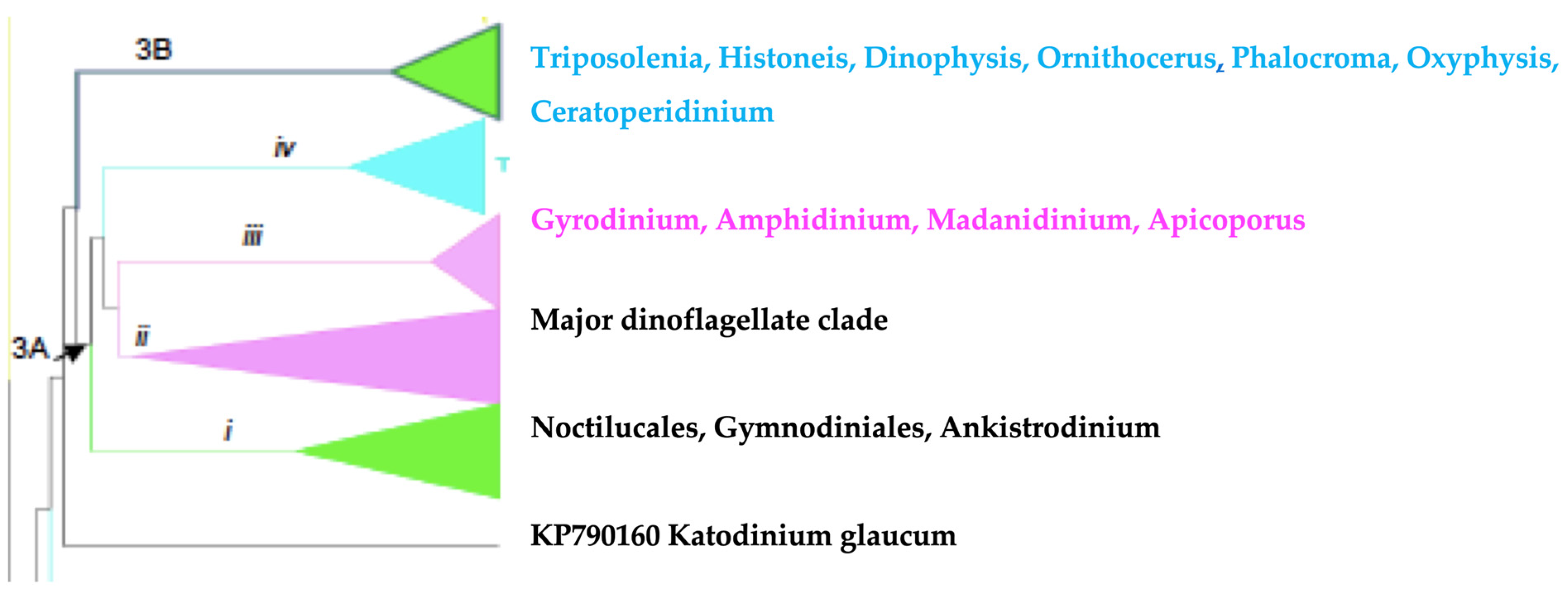
Table 1.
Comparison of features of sexual reproduction in 25 dinoflagellate species: Full references of all the authors cited in this table can be found in Bhaud et al. [12].
Table 1.
Comparison of features of sexual reproduction in 25 dinoflagellate species: Full references of all the authors cited in this table can be found in Bhaud et al. [12].
| Gametes | Zygotes | |||||||
|---|---|---|---|---|---|---|---|---|
| Species | F or M a | Different from Vegetative Cells b | Iso (+) or Aniso (-) Gamy | Naked (-) or Thecate (+) | Fertilisation Tube c | Protoplasmic Fusion d | Planozygote (Number of Flagellae) e | Hypnoyzygote f |
| Peridinium cinctum | F | + | - | - | ? | + | + 2 | + |
| F | + | - | -/+ | + | + | + 4 | + | |
| Peridinium willei | F | + | - | - | - | + | + 2 | + |
| P. gatunense | F | + | - | + | + | + | + ? | + |
| P. volzii | F | + | - | -/+ | + | + | + ? | + |
| P. limbatum | F | + | - | + | - | + | + ? | + |
| P. inconspicuum | F | + | - | + | ? | + | + ? | + - |
| P. cunningtonii | F | + | - | -/+ | - | + | + 4 | + |
| Gymnodinium excavatum | F | + | - | ? | ? | ? | + ? | + |
| G. paradoxum | F | + | - | ? | ? | ? | + ? | + |
| G.. pseudopalustre | F | + | - | ? | + | + | + 4 | + |
| Gymnodinium. breve | M | + | - | + | - | + | + 4 | - i |
| Gyrodinium uncatenatum | M | + | + | + | + | + | + 4 | + |
| Woloszynskia apiculata | F | + | - | + | + | + | + 4 | + |
| Ceratium cornutum | F | + | + | + | + g | + h | + ? | + |
| C. horridum | M | + | + | + | + g | + h | + ? | + |
| Oxyrrhis marina | M | + | - | ? | ? | ? | + ? | ? |
| Crypthecodinium cohnii | M | + | + - | ? | ? | + | + ? | + |
| M | ? | + - | + | + | + | + ? | + | |
| Gonyaulax tamarensis | M M | + + | + ? | ? ? | - ? | + ? | + 4 + ? | + + |
| G. excavata | M | ? | ? | ? | ? | ? | ? | + |
| G. monilata | M | + | - | + | - | + | + 4 | + |
| Protogonyaulax catenella | M | + | - | + | - | + | + ? | + |
| Helgolandinium subglobosum | M | + | + | ? | ? | ? | + ? | - |
| Amphidinium carteri | M | - | - | ? | + | + | + ? | + |
| Noctiluca miliaris | M | + | - | - | - | + | + 0 | ? |
| Protocentrum micans | M | - | - | + | + | - | + 2 | - |
a F, Freshwater; M, Marine water. b Differences observed between vegetative cells and gametes are often morphological variations, such as variations in cell volume and colour; gametes are generally smaller and less pigmented than vegetative cells: +, different, -, the same. c Fertilization tube or similar structure: +, present, -, absent. d Protoplasmic fusion of the two gametes: +, fusion, -, no fusion. e Planozygote: +, present, -, absent. f Hypnozygote: +, present, -, absent. g The female engulfs the male. h Protoplasm from female and male gametes is mixed.
Disclaimer/Publisher’s Note: The statements, opinions and data contained in all publications are solely those of the individual author(s) and contributor(s) and not of MDPI and/or the editor(s). MDPI and/or the editor(s) disclaim responsibility for any injury to people or property resulting from any ideas, methods, instructions or products referred to in the content. |
© 2022 by the authors. Licensee MDPI, Basel, Switzerland. This article is an open access article distributed under the terms and conditions of the Creative Commons Attribution (CC BY) license (https://creativecommons.org/licenses/by/4.0/).
Share and Cite
MDPI and ACS Style
Riding, J.B.; Fensome, R.A.; Soyer-Gobillard, M.-O.; Medlin, L.K. A Review of the Dinoflagellates and Their Evolution from Fossils to Modern. J. Mar. Sci. Eng. 2023, 11, 1. https://doi.org/10.3390/jmse11010001
AMA Style
Riding JB, Fensome RA, Soyer-Gobillard M-O, Medlin LK. A Review of the Dinoflagellates and Their Evolution from Fossils to Modern. Journal of Marine Science and Engineering. 2023; 11(1):1. https://doi.org/10.3390/jmse11010001
Chicago/Turabian StyleRiding, James B., Robert A. Fensome, Marie-Odile Soyer-Gobillard, and Linda K. Medlin. 2023. "A Review of the Dinoflagellates and Their Evolution from Fossils to Modern" Journal of Marine Science and Engineering 11, no. 1: 1. https://doi.org/10.3390/jmse11010001
Note that from the first issue of 2016, this journal uses article numbers instead of page numbers. See further details here.